Download
1 / 48
490 likes | 503 Views
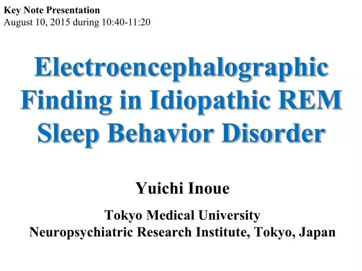
Key Note Presentation August 10, 2015 during 10:40-11:20. Electroencephalographic Finding in Idiopathic REM Sleep Behavior Disorder. Yuichi Inoue Tokyo Medical University Neuropsychiatric Research Institute, Tokyo, Japan. Conflict of Interest Disclosures – Authors/Presenters.

E N D
Key Note Presentation August 10, 2015 during 10:40-11:20 Electroencephalographic Finding in Idiopathic REM Sleep Behavior Disorder Yuichi Inoue Tokyo Medical University Neuropsychiatric Research Institute, Tokyo, Japan
Conflict of Interest Disclosures –Authors/Presenters The authors do not have any potential conflicts of interest to disclose, OR The authors wish to disclose the following potential conflicts of interest related to content in this lecture: ✔
REM sleep behavior disorder (RBD) Dream enactment behavior based on REM without atonia (kicking, punching and shouting etc), a possible cause of sleep related injury (at least 0.5%) Prevalent in elderly population Male predominance Chronic course One of prodromal symptoms of alpha-synucleinopathies JSC
JSC • Repeated episodes of sleep related vocalization and/or complex motor behaviors. • These behaviors are documented by polysomnography to occur during REM sleep or, based on clinical history of dream enactment, are presumed to occur during REM sleep. • Polysomnographic recording demonstrates REM sleep without atonia (RWA) • The disturbance is not better explained by another sleep disorder, mental disorder, medication, or substance use. REM Sleep Behavior Disorder Diagnostic Criteria Criteria A-D must be met (International Classification of Sleep Disorders Third Edition. American Academy of Sleep Medicine. 2014; pp246-247.)
JSC REM sleep PSG showing submental EMG activity (9) despite the emergence of gross arm and leg movements that are noted by the technician and reflected by prominent twitching in the upper and lower extremity EMGs (10, 11, 13, 14). REM (7, 8) immediately precedes the onset of complex behavior. F = Frontal; M = mastoid; C = central; O = occipital; LEOG/REOG = left/right electrooculogram; Ant Tib = anterior tibialis. (Inoue Y, et al. Neuropsychobiology. 2015; 71: 25–33.)
JSC REM sleep without atonia REM sleep without atonia -Phasic electromyographic (EMG) activity defined as 3-second mini-epochs including phasic EMG activity with any burst of EMG activity lasting between 0.1 and 5.0 s and an amplitude exceeding twice the background EMG activity. -Tonic EMG activity with an amplitude of at least twice the background EMG muscle tone or more than 10 μV for more than 50% of a 30-second epoch. –Both of these are mixed into normal REM episodes. In the third edition of The International Classification of Sleep Disorders (ICSD-3), any (tonic/phasic) chin EMG activity combined with bilateral phasic activity of the flexor digitorum superficialis muscles in >27% of REM sleep, scored in 30-second epochs, is recommended to distinguish RBD patients reliably from controls.
JSC (a) Appearance of stage REM and Stage RWA in the parasomnia group(a) and the control group(b). □ Stage RWA ■ Stage REM (b) The ordinate is the mean percentages of Stage REM (shaded area) and Stage RWA (unshaded area) in each 5-min sleep. Tachibana N et al. Biol Psychiatry. 1991
JSC Average REM densities in the first, Second, and third 3-hr period of nocturnal sleep in the subjects with parasomnia and the controls. (*p<0.05; **p<0.01). Tachibana N et al. Biol Psychiatry. 1991
JSC Five domain scores in NEO-PIR for groups of iRBD and HC Areas between dotted lines represent normal levels in the five domains. Patients with iRBD do not have any specific character trait. NEO-PIR, the revised version of NEO personality inventory; iRBD, idiopathic rapid eye movement sleep behavior disorder; HC, healthy control. Student t-test. (Sasai T, Inoue Y, et al. Parkinsonism Relat Disord. 2012; 18(5): 616-618.)
(Luppi PH et al. Sleep Med Rev. 2011; 15(3): 153-163) JSC Ach, acetylcholine; BF, Basal Forebrain; PH, posterior hypothalamus; SLD, sublaterodorsal nucleus; vlPAG, ventrolateral periaqueductal gray; GiV, ventral gigantocellular reticular nucleus; LC, locus coeruleus; DPGi, dorsal paragigantocellular reticular nucleus; DRN, dorsal raphe nucleus; TMN, tuberomammillary nucleus. In healthy REM sleep, the SLD–GiV circuit inhibits motoneurons, which preventspyramidal neurons in the motor cortex from producing movement. However, in patients with RBD, degeneration of the SLD–GiV circuit releases motoneurons from their normal source of inhibition, which allows excitatory projections from the motor cortex (via brainstem reticular neurons) to produce motor behaviors during REM sleep.
JSC Circuitry responsible for motor control in rapid eye movement (REM) sleep and its potential involvement in REM sleep behavior disorder (RBD). During REM sleep, the descending REM-ON glutamatergic neurons of the sublaterodorsal tegmental nucleus (SLD) excite the REM-ON GABA- and glycine-releasing neurons in the ventral gigantocellular reticular nucleus (GiV). These GiV neurons project to and inhibit skeletal motoneurons, which causes REM sleep atonia. Another population of ascending SLD neurons induce activation of the cortex during REM sleep (including the motor cortex) by exciting intralaminar thalamocortical neurons. In healthy REM sleep, the SLD–GiV circuit inhibits motoneurons, which prevents pyramidal neurons in the motor cortex from producing movement. However, in patients with RBD, degeneration of the SLD–GiV circuit releases motoneurons from their normal source of inhibition, which allows excitatory projections from the motor cortex (via brainstem reticular neurons) to produce motor behaviors during REM sleep. (Peever J et al. Trends Neurosci. 2014; 37(5): 279-88.)
JSC Statistical parametric mapping (t) axial intensity projection maps rendered onto a stereotactically normalized magnetic resonance imaging (MRI) scan, showing areas of significant increases of mean diffusivity values (color code, yellow to orange) in a cohort of patients with idiopathic rapid eye movement (REM) sleep behavior disorder versus healthy control subjects. The REM-off region is represented by the periaqueductal gray matter (PAG) in red, and the REM-on region is represented by the precoeruleus (PC) and sublaterodorsal nucleus (SLD) in green. Nuclei in yellow and blue are known to influence REM and non-REM sleep circuits. LC 5 locus coeruleus; PPN 5 pedunculopontine nucleus. Scherfler C et al. Ann Neurol. 69:400-7 (2011)
JSC Ictal SPECT of Patient RD, male with idiopathic REM sleep behaviour disorder. Difference images of cerebral perfusion, measured by Tc-99m ECD SPECT (in colour), overlaid on T1-MRI (greyscale). The colour of the SPECT indicates the degree of ictal hyperperfusion, ranging from weak (blue and green) to strong (orange and red). The arrows indicate hyperperfusion in brain regions. Bifrontal activation in premotor area, pons and anterior cerebellum. A =anterior; H =head; P =posterior; F =foot; R =right; L=left. (Mayer G et al. Brain. 2015; 138(Pt 5): 1263-1270 .)
JSC Neuropathologic findings in the locus ceruleusof a case with idiopathic RBD (A) Control: Melanized cells are not present in the locus ceruleus. Hematoxylin-eosin, X160 before 3% reduction. (B) iRBD patient: Typical eosinophilic Lewy body with a halo and core in a melanized cell was observed in the locus ceruleus. Uchiyama M et al. Neurology.1995
Risk of the development of neurodegenerative disease in idiopathic RBD patients Subject patients in = 93 ↓ 26 patients (30.0%) developed neurodegenerative disorder within 5 years (PD in =14, DLB in = 7, AD in = 4, MSA in = 1) Postuma RB, et al. Neurology 2009.
JSC Rates of neurological-disease-free survival according to the time of iRBD diagnosis ( a ) and estimated RBD onset ( b ). (Inoue Y, et al. Neuropsychobiology. 2015; 71: 25–33.)
JSC (Braak H, et al. J Neurol 2002; 249 (Suppl 3): III/ 1-III/ 5)
JSC Stage 1 : Olfactory dysfunction Autonomicdysfunction Stage 2 : REM-Sleep-behaviour disorder Stage 3 : Nigrostriatal degeneration Dopamine-Transporter-SPECT Motor symptoms Akinesia, Rigidity, Tremor ?
JSC rCBF decrease (A) and increase (B) in patients with iRBD in comparison with controls t value Vendette M et al.: Mov Disord. 2011; 26(9): 1717-24.
Biological markers of α-synucleinopathies in iRBD Mild motor symptoms Sensory disturbances Olfactory deficits Color identification deficits Autonomic dysfunction Slowing of EEG (awake and REM sleep) Cognitive deficits JSC
JSC Odor Stick Identification test for Japanease (OSIT-J) 臭素 12種類 • いおう • 墨汁 • ニス • 畳 • 分からない • 無臭
JSC Olfactory discrimination in patients with idiopathic REM sleep behavior disorder (iRBD), Parkinson’s disease (PD), and Controls. Controls Shown are scatter plots of individual scores for the odor stick identification test for Japanese (OSOT-J). Horizontal lines indicate mean levels. Miyamoto T, Hirata K et al. Mov Disord (2009)
JSC Disturbance in color identification RBD78years, male Control Fransworth-Munsell100 Hue Test Total Error Score204 Total Error Score 36
JSC ControlRBDpatient delayed image Almost no uptake Cardiac 123I-MIBG uptake in a patients with RBD Miyamoto T, Hirata K, Inoue Y et al. Neurology (2006)
JSC Cardiac 123I-MIBG scintigraphy in RDB Miyamoto T, HirataK,Inoue YNeurol(2007)
JSC Boxplot presentation of blood pressure changes measured byorthostatic standing test in controls, iRBD patients and PD patients.Systolic blood pressure (p = 0.034), Diastolic blood pressure(p = 0.017). White bars systolic blood pressure values; grey barsdiastolic blood pressure values FrauscherB, Inoue Y. et al. J Neurol. 2012; 259(6):1056-1061.
JSC Which parameter can predict the early development of α-synucleinopathies from iRBD? • Larger amount of RWA (Postuma, 2009) • Low cardiac MIBG uptake? • -Positivity is too high and not suitable for the prediction, • 3. Visa-spatial function? • 4.Olfactory dysfunction? (Mahlknecht P et al, 2015)
JSC Baseline evaluations in patients with iRBD who developed disease or remained disease-free Abbreviations: AUC = area under the curve; CI = confidence interval; UPDRS-III = motor section of Unified Parkinson’s Disease Rating Scale. The AUC values give an estimate of the diagnostic accuracy of the motor examination and olfactory testing for predicting the transition to a Lewy body disease. a Results represent means ± SD; p values calculated with an unpaired t test. b The p value calculated with a x2 test. c Results represent means (95% CIs) adjusted for age and sex; p values calculated with an analysis of covariance with age as factor and sex as covariate. (Mahlknecht P et al. Neurology. 2015; 84(7): 654-658.)
JSC Previous reports on quantitative and qualitative EEG findings in iRBD • Increased slow wave sleep and increased delta power (Massicotle-Marquez, 2005) • Negative findings for slow wave abnormality (Massicotle-Marquez, 2011) -possibly due to the inclusion of dementia or early parkinsonism in their former report • Higher theta power in frontal, temporal and occipital region while awake with a lower β power in the occipital region while awake and during REM sleep (Fantini, 2003). The dominant occipital frequency was lower in iRBD. • Suggestive of impaired cortical activation during both wakefulness and REM sleep in iRBD ⇒ Age effect should be considered…
JSC Study by Rodrigues-Brazète J et al (2013) Their study found that RBD + MCI patients had higher relative θ power in the parietal, temporal and occipital regions, and lower relative α power in the occipital region compared to RBD – MCI patients and controls. The dominant occipital frequency was also slower in RBD + MCI patients compared to controls. Moreover, RBD + MCI patients had lower relative β power in the central, parietal and temporal regions compared to controls. No between-group differences were observed between RBD – MCI and controls. These findings strongly suggested the relationship between cognitive decline and EEG slowing in patients with iRBD.
JSC Control for younger RBD (3) Clinical, polysomnographic and neuropsychological findings ・ Scores of MoCA and MMSE were lower in younger iRBD compared with controls. ・RWA measures and odor function were not different between younger and older RBD patients. (Sasai T, Inoue Y, et al. Sleep. 2013; 36(12): 1893-1899.)
JSC ● : younger RBD ▲ : older RBD ■ : control Distribution of the MoCA and MMSE scores among ages ・Age dependent decrease in the scores of MoCA and MMSE were found in all the subject groups ・The MoCA score was lower than the cutoff (25 of 26) for detecting MCI in 13 of 17 (76.5%) of the younger iRBD group, 13 of 14 (92.9%) of the older iRBD group, and 1 of 17 (5.9%) of control patients. The MMSE score was lower than the cutoff (29 of 30) for detecting MCI in 10 of 17 (58.8%) ofthe younger iRBD group, 13 of 14 (92.9%) of the older iRBD group, and 4 of 17 (23.5%) of control patients. (Sasai T, Inoue Y, et al. Sleep. 2013; 36(12): 1893-1899.)
JSC Comparison of electroencephalographic spectral power during wake, REM sleep, and NREM sleep in patients with young iRBD and controls Black bars show data for the younger rapid eye movement sleep behavior disorder group. White bars show data for the age-matched control group. *P < 0.05, values are expressed as mean ± standard deviation. (Sasai T, Inoue Y, et al. Sleep. 2013; 36(12): 1893-1899.)
JSC Partial correlation coefficients of the Montreal CognitiveAssessment score to clinical rapid eye movement sleep disorder-related variables or polysomnographic variables Partial correlation analyses controlled for age were conducted on all subject patients with iRBD (n = 31). EMG, electromyogram; n.s., not significant; RBD, rapid eye movement sleep disorder; RBDQ-JP, Japanese version of REM sleep behavior disorder questionnaire; SPT, sleep period time; SWS, slow wave sleep; TDI, threshold–discrimination–identification. (Sasai T, Inoue Y, et al. Sleep. 2013; 36(12): 1893-1899.)
JSC Partial correlation between scores of Montreal Cognitive Assessment and electroencephalogram spectral powers in respective frequency bands after controlling for age Partial correlation analyses controlled by age for all patients with rapid eye movement sleep behavior disorder (n = 31). NREM, nonrapid eye movement; n.s., not significant; REM, rapid eye movement. (Sasai T, Inoue Y, et al. Sleep. 2013; 36(12): 1893-1899.)
JSC Multiple Regression Analysis Results indicated the following multiple regression equation: MoCA score = 50.871-0.116*age-5.307*log (δ power during REM sleep in occipital regions) + 0.086*TDI score. -For this equation, the R value was 0.773, R2 was 0.598, and the regression coefficients were -0.558 for age, -0.468 for δ power, and 0.357 for TDI score (F = 9.900, P < 0.001). Electroencephalographic slowing, especially during REM sleep and olfactory dysfunction, was revealed to be associated with cognitive decline in idiopathic rapid eye movement sleep behavior disorder. (Sasai T, Inoue Y, et al. Sleep. 2013; 36(12): 1893-1899.)
JSC Controlled studies of cognitive performance in iRBD Yes = Patients show poorer performance than controls (p < 0.05); No = similar performance between patients and controls. a, b Share common participants. • MCI occurs in approximately 50% of iRBD patients (controls : 8%). • Attention, executive function, episodic verbal memory (mainly free recall capacities) and nonverbal learning are the most affected domains. (Inoue Y, et al. Neuropsychobiology. 2015; 71: 25–33.)
JSC Comparision of performance in Iowa Gambling Task performance of idiopathic RBD patients and healthy controls. (a) Total gain (point), (b) IGT score (C + D) - (A + B), (c) IGT scores for each block (C + D) - (A + B). *: p < .05, † p < .01, compared to the HC group. The boxes show mean ± SD and the error bars show the area between the lowest score and the highest score. IGT, Iowa Gambling Task; HC, healthy control; iRBD, idiopathic rapid eye movement sleep behavior disorder. Total gain and IGT score as well as IGT scores in the first, third and final banks were lower than these of the control group. (Sasai T, Inoue Y, et al. Sleep Med. 2012; 13(3): 301-306.)
JSC Hypocholinergic function? • Evidence of the involvement of the pons including PPN, laterodorsal segmental nucleus, LC and peri-LC derives from neurpathological studies on iRBD patients. • The PPN and LC representing the largest clusters of cholinergic function are known to play a role in arousal, cortical activation and cognitive function. • It is also known that cholinergic activity promotes REM sleep, and that cholinergic denervation of the limbic system is a robust determinant of hyposmia. • The short latency afferent inhibition (SAI) of the motor cortex with transcranial magnetic stimulation manifests the cholinergic function of the brain, and the SAI is clearly impaired in iRBD patients (with MCI). In these patients, SAI values are also correlated with executive functions or verbal memory (Nardone et al , 2012)
JSC Cholinergic neuronal loss in PPN/LDT in LBD. (a) Choline acetyl transferase (ChAT) staining in the pedunculopontine/laterodorsaltegmental nucleus (PPN/LDT) of normal, Alzheimer’s disease (AD), and Lewy body disease (LBD) cases without rapid eye movement sleepbehaviour disorder (RBD) (LBD NRBD) and with RBD (LBD RBD). Photos taken at \ 20. (b) Quantification of percentages of ChAT-positive neurones out of total neurones in the PPN/LDT. *LBD RBD contained significantly less ChAT-positive neurones than normals and AD. Dugger BN et al.: Neuropathol Appl Neurobiol. 2012; 38(2): 142-52.
JSC Patients with RBD + MCI had marked EEG slowing (increased delta and theta activity) in central and occipital regions during wakefulness and REM sleep, particularly in the right hemisphere, when compared with controls and, with IRBD subjects who remained idiopathic. Log-transformed spectral EEG power during REM sleep in controls (black bars) patients with IRBD (gray bars) and patients with RBD who later developed mild cognitive impairment (white bars) in C3, C4, O1 and O2 regions across delta, theta, alpha, beta1 and beta2 frequency bands. x = p < 0.05, xx = p < 0.01, xxx = p < 0.001. The EEG spectral pattern of the RBD + MCI group was similar to that seen in patients with dementia with Lewy bodies and Parkinson’s disease associated with dementia. (Iranzo A et al. Sleep Med. 2010;11(6): 534-539.)
JSC Three-dimensional views of decreased studies showed (a) regional cerebral blood flow (rCBF) at first single photon emission computed tomography (SPECT) and (b) second SPECT of rapid eye movement sleep behavior disorder (iRBD) patients compared with normal controls. Three-dimensional views of (c) decreased rCBF at second SPECT compared with first SPECT of iRBD patients. (Sakurai H, Inoue Y, et al. Geriatr Gerontol Int. 2014; 14(1): 115-120.)
JSC Conclution • EEG slowing is one of specific findings in iRBD, and this may become a candidate of the predictors of the development of neurodegeneration. • Future studies should clarify the cutoff EEG power (especially for younger patients) for the prediction. • The corresponding area of EEG abnormalities should also be investigated with neuroimaging studies especially by using acetylcholine ligands .
JSC Summary of participants. Data are expressed as mean ± SD.iRBD; idiopathic rapid eye movement behavior disorder, RBDSQ-J; Japanese version of RBD Screening Questionnairre. n.s.: Not significant. a The levels of fatigue and sleepiness before performing Iowa Gambling Task were evaluated using the visual analogue scale (0–100). (Sasai T, Inoue Y, et al. Sleep Med. 2012; 13(3): 301-306.)
JSC Comparison of EEG spectral power during wake ( a ), REM sleep ( b ) and NREM sleep ( c ) in patients with iRBD and controls. ■ = Younger RBD group; □ = age-matched control group. * p < 0.05, means ± SD. (Inoue Y, et al. Neuropsychobiology. 2015; 71: 25–33.)
JSC Diagnostic accuracy of olfactory assessment and its value for potential interventional trials in patients with iRBD Scatterplots of the identification subscore (A) and the Sniffin’ Sticks total score (B) in subjects with iRBD developing a Lewy body disease and in those remaining disease-free. Single values are given with the respective group median and the 25th and 75th percentiles. The dotted lines represent the receiver operating characteristic–based cutoff values (<7/16 for the identification subscore [A] and <18/48 for the Sniffin’ Sticks sum score [B]) and correspond also to the cutoff between the lowest and the middle tertile of olfactory function in the entire iRBD group. At these cutoffs, both tests yielded the same diagnostic accuracy of 82.4% (95% CI: 66.1%–92.0%) with a sensitivity of 77.8% (95% CI: 44.3%–94.7%), a specificity of 84.0% (95% CI: 64.7%– 94.2%), a positive predicate value of 63.6% (95% CI: 35.2%–85.0%), and a negative predictive value of 91.3% (95% CI: 72.0%–98.8%) in predicting the development of a Lewy body disease in patients with iRBD. The relative risk for a Lewy body disease in the lowest tertile of olfactory function was 7.3 (95% CI: 1.8–29.6) compared with the top 2 tertiles. Required sample sizes for randomized, placebo-controlled, 5-year follow-up interventional trials with effect sizes ranging from 20% to 50% were estimated based on the conversion rate to a Lewy body disease obtained in the total iRBD cohort (C) vs the iRBD subcohort with olfactory dysfunction (D). The green lines represent calculations at 80%, the blue lines at 90% power. (Mahlknecht P et al. Neurology. 2015; 84(7): 654-658.)