Download
1 / 59
610 likes | 890 Views
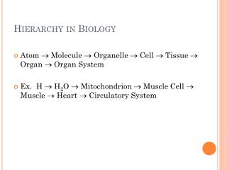
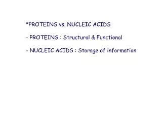
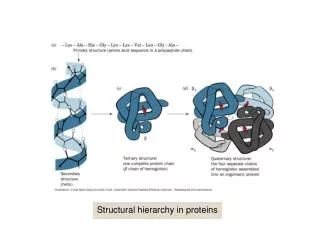
Structural hierarchy in proteins. The reaction of dansyl chloride in end group analysis. The hypothetical rate of the carboxypeptidase-catalyzed release of amino acids: all bonds cleaved at the same rate.

E N D
The hypothetical rate of the carboxypeptidase-catalyzed release of amino acids: all bonds cleaved at the same rate
The hypothetical rate of the carboxypeptidase-catalyzed release of amino acids: Ser slow, Tyr fast, and Leu intermediate
Flow diagram for polypeptide synthesis by the solid phase method
Amino acids with benzyl-protected side chains and Boc-protected a-amino groups
Amino Acid Difference Matrix for 26 Species of Cytochrome ca
Flow diagram of the chain-terminator (dideoxy) method of DNA sequencing
A portion of the output of a four reaction/one gel sequencing system
Scanning electron microscope of human erythrocytes. (a) Normal human erythrocytes revealing their biconcave disklike shape
Scanning electron microscope of human erythrocytes. (b) Sickled erythrocytes from an individual with sickle-cell anemia
A map indicating the regions of the world where malaria caused by P. falciparum was prevalent before 1930
Rate of sequence change in evolving proteins. (a) Protein evolving at random and that initially consists of 5% each of the 20 “standard” amino acid residues
Rate of sequence change in evolving proteins. (b) A protein of average amino acid composition evolving as is observed in nature
The optical alignments of human myoglobin (Mb, 153 residues) and the human hemoglobin a chain (Hba, 141 residues)
A guide to the significance of normalized alignment scores (NAS) in the comparison of peptide sequences
Use of the Needleman-Wunsch alignment algorithm [alignment of 10-residue peptide (horizontal) with 11-residue peptide (vertical)]. (a) Comparison matrix
Use of the Needleman-Wunsch alignment algorithm [alignment of 10-residue peptide (horizontal) with 11-residue peptide (vertical)]. (b) Transforming the matrix
Use of the Needleman-Wunsch alignment algorithm [alignment of 10-residue peptide (horizontal) with 11-residue peptide (vertical)]. (c) Transformed matrix
An unrooted phylogenetic tree of the five HIPIP sequences that are aligned in Fig. 7-30c
Manipulations employed in the neighbor-joining method for the construction of a phylogenetic tree
Variation in the expression of genes that encode proteins known as cyclins (Section 34-4C) in human tissues
The generation of the gas phase ions required for the mass spectrometric analysis of proteins: electrospray ionization(ESI)
The generation of the gas phase ions required for the mass spectrometric analysis of proteins: matrix-assisted laser desorption/ionization (MALDI)
The generation of the gas phase ions required for the mass spectrometric analysis of proteins: fast atom bombardment (FAB)
The ESI-MS spectrum of the 17 kD horse heart protein apomyoglobin