Download
1 / 20
320 likes | 628 Views
Optical Coherence Tomography (OCT). Gella Laxmi 2009PHXF013P. Introduction. Standard clinical. Resolution (log). Ultrasound. 100micronm. CT and MRI. 10mic. OCT. 1mic. Confocal microscopy. Penetration depth (log). 1 mm. 1 cm. 10 cm. OCT.

E N D
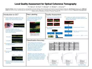
Optical Coherence Tomography (OCT) Gella Laxmi 2009PHXF013P
Standardclinical Resolution (log) Ultrasound 100micronm CT and MRI 10mic OCT 1mic Confocalmicroscopy Penetration depth (log) 1 mm 1 cm 10 cm OCT • Determining and visualizing structure that absorb and scatter light • Noninvasive in vivo analysis of retinal tissue
Principle • Michelson Interferometer
Reference beam Diode 820 Beam splitter Detector Patients eye OCT software DVD
IPL INL GCL NFL FOVEOLA OPL ONL RPE PHOTORECEPTORS ELM CHORIOCAPILLARIS Spectral Domain OCT
Features of SD-OCT • Better anatomic representation • High resolution (6 microns) • Fewer movement artifacts • Live cross-sectional movies of various details • High Signal to noise ratio • Scanning speed 25, 000 A-scans per second • 3D imaging
Retinal Structures on SD-OCT • Horizontally oriented structures – hyperreflective • Vertically oriented structures (layers containing nuclei) – hypo reflective
V V CC RPE • Choriocapillaris: • Innermost limit of the vascular layer of the eye • Thin and hyper-reflective layer • Larger vessels of choroid – hyporeflective • Inconsistently identified • Bruch’s membrane: • Not visible on SD-OCT V V CC CC RPE RPE V V
Retinal Pigment Epithelium: • RPE-CC complex divided into 3 parallel strips • 2 are thick, hyperreflective separated by thin hyporeflective line • Verhoef’s membrane
Photoreceptors: • Rods and cones contain inner and outer parts • Inner part: nuclei (outer nuclear layer) • Outer part: inner and outer segment • Connection b/w inner and outer segment forms a hyper-reflective strip (result of diff in RI) • Sharply raised at the foveola • External limiting membrane
Outer plexiform layer: • Visual cells connect to the bipolar cells • Horizontal axons of the horizontal cells • Hyper-reflective strip • Inner nuclear layer: • Nuclei of bipolar, horizontal, muller and amacrine cells • Hyporeflective layer
GCL RNFL IPL • Inner plexiform layer: • Synapses b/w ganglion cells and amacrine cells • Hyper-reflective owing to their horizontal structure • Ganglion cell layer • Bulky cells are multilayered • Hyper-reflective • Nerve fiber layer • Nerve axons • Very high reflective layer
Internal limiting membrane • Difficult to distinguish • Hyaloid and vitreous • Various pathologic structures clearly visible
Reporting SD-OCT • Comment on each layer • Reflectivity • Morphological features • Measurements of thickness
Take-home message • Retinal anatomy and virtual histology can be studied with the SD-OCT • The SD-OCT shows more detail at the vitreoretinal interface, and there is better delineation of all retinal layers
References • Bruno Lumbroso. SD-OCT Reveals Details of Posterior Segment Structures. Cataract & refractive surgery today Europe. June 2008. Pg 27-28 • Wolfgang Drexler, et al. State-of-the-art retinal optical coherence tomography. Progress in retina and eye research. 2008.Jan; 27(1): 45-88 • Bruno Lumbroso, et al. Understanding Spectral OCT. I.N.C Innovation-News-Communication. 2007. • Michael R. Hee, et al. Optical Coherence Tomography of the Human Retina. Arch Ophthalmol. 1995; 113: 325-332.