Download

1 / 51
510 likes | 767 Views
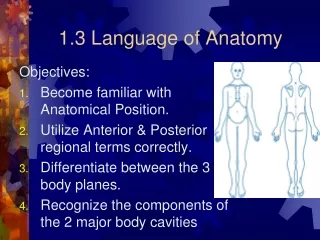
Language of Anatomy. Language of Anatomy. What is this fin called?. Language of Anatomy. Why is the back of your hand called the dorsal surface?. Language of Anatomy. To understand anatomy, common terms must be used. Anatomical Position. This defines all anatomical reference points.

E N D
Language of Anatomy What is this fin called?
Language of Anatomy Why is the back of your hand called the dorsal surface?
Language of Anatomy To understand anatomy, common terms must be used.
Anatomical Position This defines all anatomical reference points. It is defined as a person: • STANDING STRAIGHT
Anatomical Position This defines all anatomical reference points. It is defined as a person: • STANDING STRAIGHT • FACING FOREWARD
Anatomical Position This defines all anatomical reference points. It is defined as a person: • STANDING STRAIGHT • FACING FOREWARD • PALMS OUT OR FACING FORWARD
Anatomical Position This defines a persons: • Front and back also known as • Ventral and dorsal • Anterior and posterior
Anatomical Position This defines a persons: • Left versus Right
Anatomical Position This defines a persons: • Top and Bottom also known as • superior (cranial) and inferior • cephalad and caudad
Directional Terms Medial- towards the midline
Directional Terms Medial- towards the midline Lateral- away from the midline
Directional Terms Medial- towards the midline Lateral- away from the midline Intermediate-between
Directional Terms Proximal-closer to the trunk of the body
Directional Terms Proximal-closer to the trunk of the body Distal- further from the trunk of the body
Directional Terms Superficial-Towards the surface
Directional Terms Superficial-Towards the surface Deep- towards the center of the body
Body Planes &Sections Most anatomical studies, especially those involved with radiographic studies, MRI’s and CAT scans, use specific planes to study an area of the body.
Body Planes &Sections Sagittal plane-is a vertical plane that divides the body into left and right halves.
Figure 1.8c Planes of the body with corresponding magnetic resonance imaging (MRI) scans. (c) Median section (midsagittal) Median (midsagittal) plane Intestines Rectum Vertebral column
Body Planes &Sections Frontal planes-are vertical cuts that divide the body into anterior and posterior sections.
Figure 1.8a Planes of the body with corresponding magnetic resonance imaging (MRI) scans. (a) Frontal section (through torso) Left and right lungs Frontal plane Spleen Liver Heart Stomach Arm
Body Planes &Sections Transverse Planes- cut the body into inferior and superior sections
Figure 1.8b Planes of the body with corresponding magnetic resonance imaging (MRI) scans. (b) Transverse section (through torso, inferior view) Transverse plane Liver Aorta Pancreas Spinal cord Spleen Subcutaneous fat layer Body wall
Body Planes &Sections Oblique Planes- are diagonal cuts between the horizontal and vertical planes.
Body Cavities The body is divided up into two major body cavities. These are the: • Dorsal body cavity
Body Cavities The body is divided up into two major body cavities. These are the: • Dorsal body cavity and • Ventral body cavity
Body Cavities The dorsal body cavity protects the nervous system. It contains 2 subdivisions: • The cranial cavity which encases the brain
Body Cavities The dorsal body cavity protects the nervous system. It contains 2 subdivisions: • The cranial cavity which encases the brain • The vertebral or spinal cavity which encloses the spinal column
Figure 1.9a Dorsal and ventral body cavities and their subdivisions. Cranial cavity (contains brain) Thoracic cavity (contains heart and lungs) Dorsal body cavity Vertebral cavity (contains spinal cord) Diaphragm Abdominal cavity (contains digestive viscera) Pelvic cavity (contains urinary bladder, reproductive organs, and rectum) Dorsal body cavity Ventral body cavity (a) Lateral view
Body Cavities The ventral body cavity is more anterior and is the largest cavity, it contains the internal organs known as the VISCERA.
Body Cavities The ventral body cavity has two subdivisions: • The thoracic cavity
Body Cavities The ventral body cavity has two subdivisions: • The thoracic cavity and the • Abdominopelvic cavity
Figure 1.9a Dorsal and ventral body cavities and their subdivisions. Cranial cavity (contains brain) Thoracic cavity (contains heart and lungs) Dorsal body cavity Vertebral cavity (contains spinal cord) Diaphragm Abdominal cavity (contains digestive viscera) Pelvic cavity (contains urinary bladder, reproductive organs, and rectum) Dorsal body cavity Ventral body cavity (a) Lateral view
Membranes of theBody Cavities The walls of the ventral body cavities and outer surfaces of the organs are covered by a thin, double layered membrane called the Serosa.
Membranes of theBody Cavities The serosa or serous membranes are divided into 2 parts: • Parietal serosa covers the cavity walls
Membranes of theBody Cavities The serosa or serous membranes are divided into 2 parts: • Parietal serosa covers the cavity walls • Visceral serosa covers the organs
Membranes of theBody Cavities The serosa or serous membranes are divided into 2 parts: • Parietal serosa covers the cavity walls • Visceral serosa covers the organs
Membranes of theBody Cavities Together they form a potential space which has a small amount of lubricating fluid called Serous fluid.
Peritonitis Inflammation of the serous membrane, usually due to infection or trauma.
Peritonitis Inflammation of the serous membrane, usually due to infection or trauma. Leads to the organs adhering to each other, restricting movement. VERY PAINFUL
Pericarditis Inflammation of the serous membrane around the heart.
Anatomical Regions and Quadrants The Abdominopelvic Region is large and has many organ systems. It is divided up into smaller quadrants for study.
Figure 1.11 The four abdominopelvic quadrants. Right upper quadrant (RUQ) Left upper quadrant (LUQ) Right lower quadrant (RLQ) Left lower quadrant (LLQ)