Download
1 / 20
200 likes | 339 Views
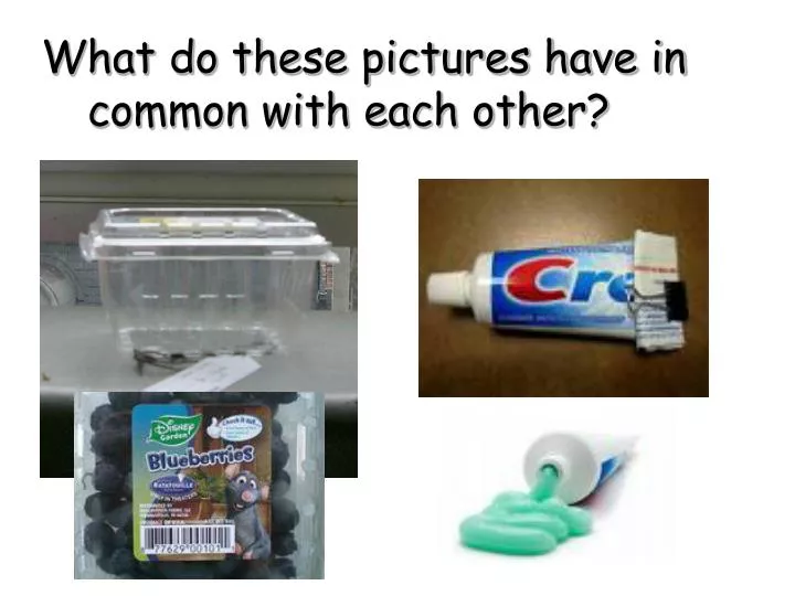
What do these pictures have in common with each other?. A thin nearly invisible structure that surrounds the cytoplasm of the cell. Continuous membrane that completely surrounds the cell. Selectively permeable. Cell Membrane. Cell Membrane; The Phospholipid Bilayer.

E N D
A thin nearly invisible structure that surrounds the cytoplasm of the cell. • Continuous membrane that completely surrounds the cell. • Selectively permeable Cell Membrane
Cell Membrane; The Phospholipid Bilayer Why do these phospholipids form this mirrored double layer? Hint…..the cell is made of about 80% water
Hydrophilic = Water loving Cell Membrane; The Phospholipid Bilayer Phospholipid ‘head’ Hydrophobic = Water fearing Phospholipid ‘tail’
Rank the molecules according to their speed to cross the cell membrane from fastest to slowest…..(or maybe not at all….)
Rank the molecules according to their speed to cross the cell membrane from fastest to slowest…..(or maybe not at all….) Nonpolar Polar Glucose (too big & polar) Ions (have a charge)
Cell MembraneFluid Mosaic Model This currently accepted model was proposed by S.Singer & G. Nicolson in 1972. The phospholipids are the round yellow structures with the blue tails, The proteins are the blue lumpy structures that are scattered around among the phospholipids. cell_membrane_transport
On your paper, read the descriptions of each part of the cell membrane & label the diagram. Carbohydrates Phospholipids Cholesterol Cytoskeleton Protein
Phospholipids • Protein – helps regulate what passes through the cell • Cholesterol – provides strength in membrane • Carbohydrates- act as source of protection & recognition of other molecules • Cytoskeleton- helps provide structure & shape to cell Cell Membrane’s Parts
Cell Membrane’s Proteins • Floating around in the cell membrane are different kinds of proteins. • They are not held in any fixed pattern but instead float around in the phospholipid layer. • Generally these proteins structurally fall into four categories...
4 Types of Cellular Membrane Proteins Enzyme proteins catalyze many chemical reactions Ex. produces cell energy molecules & perform photosynthesis in plants 1. Recognition proteins that identify the cell to other cellsEx. Immune system & blood types 2. Transport / carrier proteins that regulate molecules crossing the membrane. Ex. Ions such as Na+ or K+ or larger molecules C6H12O6 3.
4 Types of Cellular Membrane Proteins Cell Signaling allows cells to signal. Insulin is the chemical signal that operates gated channels found on the surface of muscle and liver cells. These gated channels are transport proteins that allow for the inward passage of sugar from the blood and body fluids into a cell's cytoplasm. The gated transport proteins are open for sugar transport only when signaled by the presence of insulin. 4.
Lipid Rafts ? • In artificial membranes, different lipids separate from each other based on their physical properties, forming small islands called lipid rafts. • These rafts have a higher concentration of certain specialized lipids and are also distinguished by a different assortment of proteins. Certain types of proteins cluster together in rafts, while others remain mostly outside of rafts. The big question is, to what extent do these rafts, seen readily in artificial membranes, actually exist in living cells? • Using advanced laboratory methods and imaging techniques, some researchers found evidence that rafts, indeed, do form in living cellular membranes, but these rafts may be small and transitory. Although the existence of lipid rafts in cellular membranes remains controversial, many scientists believe they serve as communication hubs by recruiting proteins that need to come together in order to transmit a signal. Researchers are beginning to link lipid rafts with a variety of diseases, including AIDS, Alzheimer's, anthrax, and atherosclerosis.
Red Blood Cells…. Some proteins on the RBC…….
Red Blood Cells…. The same thing applies with Rh factors… If a person is Rh positive, then the person has Rh antigens. If a person is Rh negative, then the person has NO antigens, and then will have Rh antibodies.