Download
1 / 44
450 likes | 542 Views
Lecture IX. Brain Pathways: Sensation. Bio 3411 Wednesday September 23, 2009. What the last lecture was about Spinal Cord Peripheral Nerves Spinal Nerves Spinal Reflexes Introduction to Pathways. “Readings” (background only). Neuroscience 4 th ed Page(s ) Feature 207 Touch

E N D
Lecture IX. Brain Pathways: Sensation Bio 3411 Wednesday September 23, 2009
What the last lecture was about Spinal Cord Peripheral Nerves Spinal Nerves Spinal Reflexes Introduction to Pathways
“Readings”(background only) Neuroscience 4thed Page(s) Feature 207 Touch 231 Pain 253 Eye 289 Central Visual Pathways 313 Auditory Function 343 Vestibular Function 363 Olfactory receptors 381 Taste buds & receptors The Brain Atlas 3rd ed Page(s) Feature 182-183 Dorsal Column/Medial Leminscus – Touch & Position 186-187 Spinothalamic Tract – Crude touch, Pain & Temperature 41, 45 Brain stem showing optic pathway 192-193 Visual Pathways 196-197 Auditory Pathways 198-199 Vestibular Pathways 194-195 Olfactory Pathways 190-191 Taste Pathways Lecture IX. Brain Pathways: Sensation
References †Cox, J. J., Reimann, F., Nicholas, A. K., Thornton, G., Roberts, E., Springell, K., Karbani, G., Jafri, H., Mannan, J., Raashid, Y., Al-Gazali, L., Hamamy, H., Valente, E. M., Gorman, S., Williams, R., McHale, D. P., Wood, J. N., Gribble, F. M., & Woods, C. G. (2006). An SCN9A channelopathy causes congenital inability to experience pain. Nature, 444(7121), 894-898. †Goodwin, G. M., McCloskey, D. I., & Matthews, P. B. (1972). Proprioceptive illusions induced by muscle vibration: contribution by muscle spindles to perception? Science, 175(28), 1382-1384. †Mogil, J. S., Yu, L., & Basbaum, A. I. (2000). Pain genes?: natural variation and transgenic mutants. Annu Rev Neurosci, 23, 777-811. _________________ † Posted on class web site. Lecture IX. Brain Pathways: Sensation
What the last lecture was about: Spinal Cord - Segmental organization Peripheral Nerves - Compound action potential (Erlanger & Gasser Prix Nobel 1944) Spinal Nerves - Dermatomes, motor neuron pools (nuclei) and motor units Spinal reflexes - stretch (knee jerk); withdrawal/ crossed extensor Introduction to Pathways - 1 sensory (DC-ML); 1 motor (CST) Lecture VIII. The Spinal Cord, Relexes and Brain Pathways
Overview Sensation Sensory Transduction Receptive Fields Adaptation Feature Detection Maps Lecture IX. Brain Pathways: Sensation
Charles Scott Sherrington 1857 – 1957 (Prix Nobel 1932) Edgar Douglas Adrian 1889 – 1977 (Prix Nobel 1932) Lecture IX. Brain Pathways: Sensation
The Five Senses • Touch: e.g., fine, muscle/position, pain • Smell: e.g., odorants, “taste”, opposite sex • Taste: bitter, sweet, sour, salt, ?(glutamate/umami) • Hearing/Balance: e.g., frequency & amplitude; linear & angular acceleration • Sight: light/dark, color (chromatic) Lecture IX. Brain Pathways: Sensation
Domains • Exteroception vs Interoception • Distance vs Direct Lecture IX. Brain Pathways: Sensation
Sensory Transduction • Single fiber recording (E. Adrian, Prix Nobel 1932) • Transduction is the conversion of a relevant physical stimulus into altered membrane potential, the currency of the nervous system. • Stimuli: • radiant – light and thermal • mechanical – pressure and sound • chemical – molecules and ions Lecture IX. Brain Pathways: Sensation
Na+ Na+ Na+ Na+ Na+ Na+ Na+ Na+ General Scheme for Sensory Transduction Interaction with Cell Stimulus Conductance Change Receptor Potential Neural Activation Lecture IX. Brain Pathways: Sensation
Transducers • Direct: by neurons • Mediated: by extensions, cell filters, receptor cells, complex organs • Code: onset, duration, intensity, change Lecture IX. Brain Pathways: Sensation
Touch “Receptors” in Skin There are many different kinds of sensory endings in the skin. They are relatively more sensitive to movement (amplified by the lever of a hair (B)), vibration, light pressure, pain, temperature etc. Lecture IX. Brain Pathways: Sensation
Photoreceptors in Eye - Sight The sensors in the eye contain “visual pigments” that change chemically when exposed to light of different colors and intensities. Photoreceptors sensitive to red, blue and green are called cones (C) while those sensitive to low light levels are rod like (R). Lecture IX. Brain Pathways: Sensation
Hair Cells in Ear The sensors in the ear are modified “touch” receptors. Sound causes the membrane on which these “hair cells” (because they have cell protrusions that look like hairs) rest to move and this causes the hairs to bend. When the hairs bend the hair cells depolarize and release transmitter to activate the sensory nerve endings. Lecture IX. Brain Pathways: Sensation
With respect to neurons: • Threshold (the magnitude of a stimulus sufficient to depolarize the sensory neuron) • Adequate Stimulus (the form of energy to which a particular sensory cell is most sensitive - light, touch, sound, etc.) • Law of specific nerve energies (depolarization of neurons in a pathway is interpreted as a particular form of stimulation - pressure to the eyes or direct electrical activation of the visual cortex are both interpreted as a change in light) Lecture IX. Brain Pathways: Sensation
Thresholds by Location The threshold to pressure differs over the body. The lips and the ends of the fingers are most sensitive. In part, this reflects different innervation densities (higher in the fingers and lips). Similar differences innervation density associated with high acuity vision and speech sounds are found in the eye and the ear respectively. Lecture IX. Brain Pathways: Sensation
Thresholds by Fiber Type The thresholds to mechanical force (mbars) differ for endings associated with different fiber sizes. Smaller forces activate myelinated faster conducting fibers (A - b) while greater forces are required to activate unmyelinated C and thin myelinated slower conducting fibers (A -d). Lecture IX. Brain Pathways: Sensation
Receptive Fields • Mainly about change • Tuning and fidelity • Organization - orientation, direction Lecture IX. Brain Pathways: Sensation
Receptive Fields - Endings Single sensory neurons innervating the hand have different receptive fields depending on the kind of ending they are associated with. These different endings (here named for famous guys in Italy or Germany) respond to very localized stimulation (Meissner & Merkel) or to more widely placed stimuli (Pacini and Ruffini). Lecture IX. Brain Pathways: Sensation
Receptive Fields - Retina (Center/Surround) The neurons projecting from the eye to the rest of the brain (ganglion cells) respond stimuli in the center of their receptive fields by increasing depolarization (which will increase firing) while stimuli in the periphery of the receptive field will hyperpolarize them (which will make the cell less likely to fire). The cell fires best when the stimulus covers only the central excitatory part of the receptive field as shown in the histogram at the bottom. Lecture IX. Brain Pathways: Sensation
Receptive Fields - Visual Cortex (Orientation & Length) A neuron in the visual cortex that responds best to stimuli of a particular lengths, in a particular orientation, moving in a particular direction at a prefered speed. (The bar in A is the right length. The one in B is too long and the cell fires less.) Lecture IX. Brain Pathways: Sensation
General Scheme for Neuron “Adaptation” Sensory Neuron Rapidly Adapting Rapidly/ Slowly Adapting Slowly Adapting Lecture IX. Brain Pathways: Sensation
Maps • Somatotopic, Visuotopic, Tonotopic, etc. • All Levels • Distortions ≈ innervation density Lecture IX. Brain Pathways: Sensation
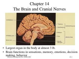
Dorsal Column – Medial Lemniscus Pathway This pathway carries fine discriminative and active touch, body and joint position, and vibration sense.
THE BRAIN ATLAS, 3rd ed, p 185 Face Hand Body Foot Lecture IX. Brain Pathways: Sensation
This is a sketch of the left cerebral hemisphere of a monkey brain. The body parts to which neurons in the cerebral cortex of the monkey best respond are organized in 2 systematic maps (Sm I and Sm II) in the parietal lobe. Lecture IX. Brain Pathways: Sensation
The whiskers on the mouse’s face are innervated by sensory neurons that project to a pathway ending in the somatosensory cortex. In sections parallel to the surface of the cortex, simple stains show a “visible” map of the whiskers and easily identify groups of cells which fire when the homologous whisker is moved. Lecture IX. Brain Pathways: Sensation
Visual Pathways These pathways convey visual information for recognizing scenes and objects, directing gaze, controlling light levels on the retina, and modulating body function with changes in the length of the day.
Eye Optic nerve Hypothalamus Optic chiasm Optic Tract Pretectum Lateral geniculate nucleus Superior Colliculus (Optic Tectum) Optic Radiation Visual Cortex THE BRAIN ATLAS, 3rd ed, pp. 192-193 Lecture IX. Brain Pathways: Sensation
Innervation of Visual Cortex from One Eye (via LGN) The axons to the visual cortex of monkeys that represent one eye are separate from those from the other eye. A technique was used that labeled axons from one eye. The image above cuts through the thickness of the visual cortex showing patches; the one below was reconstructed from sections cut in the plane of the cortex showing that the patches above are actually stripes (ocular dominance stripes). Lecture IX. Brain Pathways: Sensation
The map of the visual world (right) onto the visual cortex of the monkey (dark area in the box to the left). Lecture IX. Brain Pathways: Sensation
Orientation Map in the Monkey Visual Cortex (Optical Imaging) The different colors represent areas responding to bars of light in different parts of the visual field in different orientations as indicated in the key on the left of the figure. Lecture IX. Brain Pathways: Sensation
The 6th Sense Lecture IX. Brain Pathways: Sensation
Motor Unit - A motor neuron and the muscle fibers it innervates. Lecture VIII. The Spinal Cord, Relexes and Brain Pathways
Demonstration Lecture IX. Brain Pathways: Sensation
Loss of myelin in dorsal columns in late stage Syphilis – Tabes Dorsalis Lecture IX. Brain Pathways: Sensation
Two Clinical Findings in • Tabes Dorsalis: • Difficulty walking over uneven surfaces in the dark. • Reduced or absent knee jerk reflexes. Lecture IX. Brain Pathways: Sensation
What this Lecture was About Sensory Transduction Receptive Fields Adaptation Feature Detection Maps Sensory Integration Lecture IX. Brain Pathways: Sensation