Download
1 / 82
820 likes | 1.07k Views
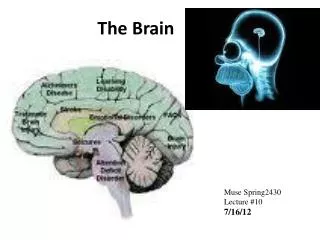
Traumatic Brain Injury & Brain Tumors Fall 2011. John Nation, RN, MSN Adapted from the notes of Marnie Quick, RN, MSN and Charlene Morris, RN, MSN. The Brain. Brain A & P. Three major components- Cerebrum Right and left hemispheres Four lobes- frontal, temporal, parietal, and occipital

E N D
Traumatic Brain Injury&Brain TumorsFall 2011 John Nation, RN, MSN Adapted from the notes of Marnie Quick, RN, MSN and Charlene Morris, RN, MSN
Brain A & P Three major components- Cerebrum • Right and left hemispheres • Four lobes- frontal, temporal, parietal, and occipital Frontal Lobe- • Cognitive function • Memory retention • Voluntary eye and motor movements • Expressive speech via Broca’s area Temporal Lobe- • Speech reception via Wernicke’s area • Visual and auditory integration Parietal Lobe- • Spatial information and control Occipital Lobe- • processing of sight
Brain A & P (Cont’d) Brainstem- • Includes midbrain, pons, and medulla • Respiratory function • Vasomotor function • Cardiac function • Centers for sneezing, coughing, vomiting, sucking, swallowing, and hicupping
Brain A & P (Cont’d) Cerebellum- • Located under the occipital lobe of the cerebrum • Coordinates voluntary movement • Trunk stability • Equilibrium
Incidence of Traumatic Brain Injury • 1.1 million people treated and released annually in the US • 50,000 people die and 235,000 are hospitalized • 22% of hospitalized clients die • Men twice as likely to have TBI as women
Incidence (Cont’d) • Mortality rate after severe head injury around 35% • More than half of people who survive severe head injury have serious disability
Risks • Motor Vehicle Accidents (MVA) - 42,642 deaths due to MVA in 2006 - 3,475 deaths in Texas alone • Elevated Blood Alcohol Levels • Sports-related Trauma • Recreational Activities- Cliff jumping, rock climbing, mountain biking, etc. • Assault- Firearms, blunt trauma
Mechanisms of Injury • Acceleration Injury- struck in head by a moving object • Deceleration Injury- head hit non-moving object • Blunt or penetrating injury to the brain • Closed head injury
Mechanisms of Injury (Cont’d) • Coup-countercoup injury- brain rebounds within skull, causing injuries at site of impact (coup) and directly opposite (countercoup) • Countercoup injury often more severe Source: Patrick J. Lynch, Creative Commons License 2006
Types of Head Injury Scalp Lacerations- - external - extensive bleeding secondary to lots of blood vessels with poor constrictive abilities - care focused on preventing blood loss and infection
Skull Fractures Linear Skull Fracture- • Break in continuity of bone • No alteration of relationships of parts • Frequently associated with low- speed injuries • Dura is intact • Accounts for around 80% of all skull fractures
Skull Fractures (Cont’d) Depressed Skull Fracture- • Inward indentation of skull • Frequently associated with powerful blow/ mechanism of injury
Skull Fractures (Cont’d) Simple Skull Fractures- • No fragmentation or communicating lacerations • Associated with low to moderate impact Comminuted Skull Fractures- • Multiple fractures with bone fragmentation • Associated with direct, high-momentum impact Compound Skull Fractures- • Scalp laceration and depressed fracture with communicating pathway to intracranial cavity
Basilar Skull Fractures • Fractures along the base of the skull • Can include cranial nerve deficits *Battle’s sign* *Raccoon Eyes*
Battle’s Sign (Postauricular ecchymosis) Raccoon Eyes (Periorbital ecchymosis)
Basilar Skull Fracture (Cont’d) Cerebrospinal Fluid (CSF)- • Rhinorrhea- leaking of CSF from the nose • Otorrhea- leaking of CSF from the ear • Client is at high risk for meningitis
Assessment for CSF • Test fluid for positive glucose reading- CSF gives positive glucose reading • If blood is also in CSF, testing for glucose is not reliable since blood has glucose as well • Allow fluid to leak onto 4x4, observe drainage over a few minutes, a yellowish ring around the blood indicates presence of CSF.
Complications of Skull Fracture • Intracranial infections • Hematoma • Meningeal and brain tissue damage
Types of Skull Fractures (Cont’d) • Open Skull Fracture- extension of the fracture into the dura or air sinuses • Closed Skull Fracture- can lead to increased risk for elevated ICP
Minor Brain Injury • Concussion- • Brief disruption in level of consciousness (LOC) • Retrograde amnesia (difficulty remembering event) • Headache • Usually of short duration • Usually not admitted to hospital if loss of consciousness < 5 minutes
Minor Brain Injury (Cont’d) • Concussion Precautions: Monitor for: • worsening headache • Vomiting • Confusion/ change in level of consciousness • Weakness on one side of body • Inability to wake up
Minor Brain Injury (Cont’d) Post Concussion Syndrome: • Generally 2 weeks to 2 months post injury • Headache, decreased attention span, personality/ behavioral changes, decreased short term memory, lethargy • Can significantly impact daily life
Major Head Trauma Contusion- • Bruising of the brain within an area of the brain • Usually connected with a closed head injury • May involve bleeding, necrosis, and infarction at the site of a fracture • Clients on anticoagulation therapy with contusion are at higher risk for severe injury and death • Seizure is a common complication
Major Brain Injury (Cont’d) Laceration- • Tearing of the brain tissue • Commonly seen with penetrating injuries (ie gunshot, knife wounds), open fractures, and depressed fractures • Surgical repair not possible • The larger the area of injury, the worse the outcome
Major Brain Injury (Cont’d) Diffuse Axonal Injury (DAI)- • Widespread axonal damage • Can be present with mild to severe TBI • Results in axon swelling and disconnection • Results in decreased LOC, increased ICP, and global cerebral edema • 90% of clients remain in persistent vegetative state
Major Brain Injury (Cont’d) Epidural Hematoma- • Bleeding between the dura and the skull • Is a true emergency • Usually seen with laceration to an artery, frequently the middle meningeal artery • Frequently initial loss of consciousness followed by a temporary improvement in condition (called a lucid interval)
Epidural Hematoma (Cont’d) Signs and Symptoms- • Lucid interval following initial loss of consciousness • Decreased level of consciousness • Headache • Nausea and vomiting • Progresses rapidly Rapid surgical intervention needed!
Subdural Hematoma Subdural Hematoma- • Bleed between the outer arachnoid membrane and the dura mater • Tends to be caused by a venous bleed, though can be arterial in nature • More common than epidural hematoma • Usually slower to develop than epidural hematoma
Subdural Hematoma Signs and Symptoms- • Similar to signs/symptoms of increased ICP • Decreasing LOC • Headache
Intracerebral Hematoma Intracerebral hematoma- • Caused by bleeding within the parenchyma (aka the nervous tissue) • Occurs with around 16% of head injuries
Review • Scalp lacerations • Skull fractures • Concussions • Contusions • Diffuse Axonal Injury • Epidural Hematoma • Subdural Hematoma • Intracerebral Hematoma
Collaborative Care for TBI Diagnostic Studies- • CT scan - best diagnostic study to evaluate head trauma related to rapid diagnosis and treatment • Spinal x-ray, skull x-ray • MRI • PET (Positron Emission Tomography) • Transcranial Doppler (to measure CSF velocity)
Collaborative Care for TBI (Cont’d) Diagnostic Tests (Cont’d)- • ABG’s • CBC • Glucose • Electrolytes
Assessment Findings(Pg 1484 in Lewis) • Fractures/depressions • Battle’s Sign, Raccoon eyes • Cheyne-strokes respirations • Decreased O2 saturation • Pulmonary edema
Assessment Findings (Cont’d) • Unequal/ dilated pupils • Confusion • Abusive/ slurred speech • Altered LOC • Seizures • Incontinence • Posturing • Glasgow coma scale <12 • CSF leaking form ears or nose
Initial Interventions • Ensure patent airway • Stabilize cervical spine • O2 via non-rebreather • Large bore IV access (2) • Control external bleeding • Ongoing assessment
Ongoing Monitoring (Lewis Pg. 1484) • Maintain client warmth • Frequent vital signs, level of consiousnes, SpO2, heart rhythm, Glasgow Coma Scale, pupil size/reactivity • Assess need for intubation if unable to protect own airway (absent gag reflex) • Use caution with IV fluids to avoid overload/ increasing ICP
Treatment of TBI • Prevent secondary injury by managing increased ICP • Primary treatment is quick diagnosis and surgery if necessary • In concussion and contusion, usually treated only with observation and management of increased ICP
Managing Increased ICP(At a glance from Module 10) • Airway management • Elevating head of bed 30 degrees • Temperature regulation • Osmotic diuretics (Mannitol) • Lasix • Invasive monitoring • Surgery
Prevention of Complications • Medications to prevent increased ICP • Prevent/ treat seizures • Stool softeners to prevent straining • Antipyretics, cooling • TPN, tube feedings, supplements • If CSF leak, do not pack nose/ears, no suctioning of nose, no blowing/ sniffing of nose. Lightly cover, change PRN,
Surgery for TBI In depressed and comminuted fractures, frequently a craniotomy to remove fragments • Crainiotomy- opening into the cranium with removal of a bone flap and opening the dura If large amount of bone is destroyed, may need: • craniectomy- removal of bone to allow brain to expand • Cranioplasty- repair of a cranial defect using articifial material
Surgery for TBI (Cont’d) Acute Subdural and Epidural Hematomas- • Burr-hole openings for rapid decompression • Craniotomy • Drain generally left in place post surgery to prevent reaccumulation of blood
Nursing Assessment- Review • Frequent Nuero assessments • Frequent vital signs • Glasgow Coma Scale • Brainstem reflexes (pupils, cough, gag, Doll’s eyes) • Watch for increased BP, decreased HR, and altered respirations - Cushing’s Triad- late sign with increased ICP- very bad! • Watch for CSF leak
Nursing Diagnosis • Ineffective airway clearance • Ineffective breathing pattern • Ineffective tissue perfusion • Acute pain • Anxiety • Hyperthermia • Decreased cerebral perfusion