Download
1 / 24
240 likes | 383 Views
Biomedical Engineering. Tissue Engineering of the Kidney: Cell Culture Model of the Glomerular Capillary Wall. Ian Flaherty and Nicholas Fountoulakis Technical Advisor: Dr. Joel Henderson, Boston Medical Center. Background and Motivation. Background: The Kidneys.

E N D
Biomedical Engineering Tissue Engineering of the Kidney: Cell Culture Model of the Glomerular Capillary Wall Ian Flaherty and Nicholas Fountoulakis Technical Advisor: Dr. Joel Henderson, Boston Medical Center
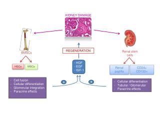
Background: The Kidneys • The kidneys provide homeostatic functions to the body and serve as a filter for blood circulating through the body • The basic unit of the kidney is the nephron (a tangle of blood vessels and tubules) • Contained in the nephron is a structure called the glomerulus which plays a major role in blood filtration
Background: The Glomerulus • The glomerulus is a tuft of capillaries found in Bowman’s capsule in the Kidney, situated between two arterioles. • It serves as the primary site of filtration in the kidney. • We are interested in the components that comprise the glomerular capillary loop.
Background: Cell Components of the Glomerulus Electron Micrograph of Glomerular Capillary Wall • Structure of the capillary wall is very unique and complex. • Three main components: endothelial cells, basement membrane, epithelial cells (podocytes). • In many disease states such as glomerulosclerosis, kidney failure is characterized as loss of protein in the urine. • The filtration barrier becomes compromised. Bowman’s Space Farquhar, M.G 2006
Motivation • Approximately 23 million Americans have physiological evidence of chronic kidney disease. • The glomerulus is commonly the site of injury in these diseases. • Need for a versatile, in vitro, model of studying glomerular function where parameters are easily varied • Potential for analyzing: • drug delivery phenomenon • genetically altered podocytes • relating permeability changes to disease states in the kidney.
Methods: Overview • Cell Culture • Device Design and Construction • Fick’s Law: Mass Flux • Microscopy • (SEM, Brightfield)
Methods: Cell Culture • Polytetrafluoroethylene (PTFE) membrane inserts were obtained from Millipore and coated with Type I rat-tail collagen. • Podocytes were cultured on the apical (top) side of the membrane.
Chamber A Chamber B Methods: Device Design and Experimental Setup Sampling Port B Sampling Port A
Methods: Permeability to Macromolecules Fick’s First Law: Js - the solute flux (mg/sec) A – the membrane’s surface area (4.2 cm2) D - the effective diffusion coefficient (cm2/s) ∆C - the concentration difference across the membrane (mg/ml) ∆x– the thickness of the membrane (30 µm) P – the effective permeability coefficient (cm/s)
Results: Morphology Comparisons • Moin Saleem Normal Human Podocytes on polystyrene (magnification unknown) • Normal Human Podocytes on PTFE (100x)
Results: SEM (Collagen Coating) Blank PTFE Membrane Membrane After Collagen Treatment
Results: Macromolecular Transport Initial [Albumin] = 0 mg/ml Final [Albumin] = 0.5 mg/ml Initial [Albumin] = 0 mg/ml Final [Albumin] = 0.16 mg/ml
Results: Macromolecular Transport • Membranes with cells allowed less mass flux at higher concentration gradients. • Concentration gradients were the only driving force for mass flow.
Discussion • Podocytes were successfully cultured in a monolayer on PTFE membranes • Comparatively, podocytes showed proper morphology to those seen in previous literature • A device was designed and built to perform perfusion experiments to “synthetic tissue” under static pressure conditions • Podocytes were shown to decrease the effective permeability of albumin through the membrane • Successfully created in vitro model to study permeability of cultured podocytes
Future Work • Establish a co-culture on PTFE membrane with podocytes and endothelial cells • Allow cells to become differentiated before perfusion • Redesign device to allow for applied pressure gradient across tissue • Knockout genes to relate changes in permeability to protein expression
References • Jennette, J.C. et al, Hepinstall’s Pathology of the Kidney. Lippincott Williams & Wilkins; Fifth edition. 20-42, 1998. • Abrahamson, D.R., Structure and Development of the Glomerular Capillary Wall and Basement Membrane. American Journal of Physiology: Renal and Fluid Electrolyte Physiology. 253:F783-F794, 1987. • Farquhar, M.G. Glomerular Basement Membrane: Not Gone, Just Forgotten. Journal of Clinical Investigation. 116:2090-2093, 2006. • Abrahamson, D. R. Origin of the Glomerular Basement Membrane Visualized After in vivo Labeling of Laminin in newborn Rat Kidneys. The Journal of Cell Biology. 100:1988-2000, 1985. • Sariola, H et al, Dual Origin of Glomerular Basement Membrane. Developmental Biology. 101:86-96, 1984. • Kanawal, Y.S. De Novo Cellular Synthesis of Sulfated Proteoglycans of the Developing Renal Glomerulus in vivo. Proceedings of the National Academy of Science. 81:7108-7111, 1984. • Hirschberg, R., S. Wang, G. M. Mitu. Functional Symbiosis Between Endothelium and Epithelium cells in glomeruli. Cell and Tissue Research. 331:485-493, 2008. • Brenner, B.M, J.L. Troy, T. M. Daugharty. Dynamics of Glomerular Ultrafiltration in the Rat. Journal of Clinical Investigation. 50: 1776-1780, 1971. • Daniels, B.S. et al. Glomerular Basement Membrane: in vitro Studies of Water Permeability. American Journal of Physiology. 262: F919-F926, 1992. • Caulfield, J.P, M.G. Farquhar. The Permeability of Glomerular Capillaries to Graded Dextrans. The Journal of Cell Biology. 63:883-903, 1974. • Farquhar, M.G. The Primary Glomerular Filtration Barrier – Basement Membrane or Epithelial Slits?. Kidney International. 8:197-211, 1975.
References cont. • Change, R.L.S. et al. Permselectivity of Glomerular Capillary Wall: III Restricted Transport of Polyanions. Kidney International, 8:212-218, 1975. • Du Bois, R., E. Stoupel. Permeability of Artifical Membranes to a Pluridisperse Solution of 251-Polyvinylpyrrolidone. Biophysical Journal. 16:1427-1445, 1976. • Tencer, J. et al. Proteinuria Selectivity Index Based Upon 2-macroglobulin or IgM is Superior to the IgG Based Index in Differentiating Glomerular Diseases. Kidney International. 54:2098-2105, 1998. • Klaentschi, K. et al. Pressure-Permeability Measurements in the Basement Membrane: Effects of Static and Dynamic Pressures. American Journal of Physiology: Heart and Circulatory Physiology. 274:1327-1334, 1998. • http://www. carolguze. com/text/102-23-Excretory%20System. shtml (nephron image reprint) • Mundel P. Rearrangements of the Cytoskeleton and Cell Contacts Induce Process Formation During Differentiation of Conditionally Immortalized Mouse Podocyte Cell Lines. Experimental Cell Research. 236:248-58, 1997. • Ballermann, B. J. Regulation of Bovine Glomerular Endothelial Cell Growth in vitro. American Journal of Physiology 256:182–189, 1989. • Eng, E. , B. Ballermann. Diminished NF-_B Activation and PDGF-B Expression in Glomerular Endothelial Cells Subjected to Chronic Shear Stress. Journal of Microvascular Research. 65:137-144, 2003. • http://www. millipore. com/images/xl/MB3973-06[757-ALL]. jpg (standing membrane insert image reprint). • Saleem MA, O'Hare MJ, Reiser J, Coward RJ, Inward CD, Farren T, Xing CY, Ni L, Mathieson PW, Mundel P. A conditionally immortalized human podocyte cell line demonstrating nephrin and podocin expression. J Am Soc Nephrol 2002 13:630-638. • O'Hare MJ, Bond J, Clarke C, Takeuchi Y, Atherton AJ, Berry C, Moody J, Silver AR, Davies DC, Alsop AE, Neville AM, Jat PS. Conditional immortalization of freshly isolated human mammary fibroblasts and endothelial cells. Proc Natl Acad Sci USA 2001 98:646-651.
Acknowledgements Faculty: • Professor Joel Henderson • Professor Joe Tien • Professor Cassandra Smith • Additional: • He Fangfang • Philip Bondzie • FelitaAgus • Hui Chen • Julie Tomolonis • Dan Flaherty • Laboratory: • Renal Pathology at Boston Medical Center