Download

1 / 27
270 likes | 433 Views
A View of the Cell. Dr. Stafford 2016. A View of the Cell. Before microscopes people believed in curses and the supernatural as the cause of diseases Microscopes opened a whole new world – Microorganisms – organisms only seen by microscope Cells – the building blocks of all living organisms.

E N D
A View of the Cell Dr. Stafford 2016
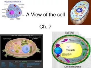
A View of the Cell • Before microscopes people believed in curses and the supernatural as the cause of diseases • Microscopes opened a whole new world – Microorganisms – organisms only seen by microscope • Cells – the building blocks of all living organisms
A View of the Cell • Anton van Leeuwenhoek used a simple microscope with natural light in mid 1600’s • Compound light microscope – 2 or more lens magnify up to 1500X’s • Had to learn size relationships • http://www.cellsalive.com
A View of the Cell • The cell theory - three main ideas 1. All organisms are composed of one or more cells – unicellular or multicellular 2. The cell is the basic unit of organization 3. All cells come from preexisting cells – cells that are living
A View of the Cell • Cell theory (cont.) • Scientists involved in its development 1. Robert Hooke – cork cells – named cells after rooms in monastery 2. Matthias Schleiden – observed plants and determined they were cells 3. Theodore Schwann – observed animals
A View of the Cell • Electron microscopes – 1940’s – 500,000X 1. Used beam of electrons instead of light 2. Scanning electron microscope (SEM) – scan surface – 3-D shape 3. Transmission electron microscope (TEM) – structures inside cell
A View of the Cell • Two different types of cells 1. Prokaryotes – means Pro = before and Karyote = nucleus – so prokaryote means cells without a nucleus. Also has no membrane- bound organelles – example bacteria
A View of the Cell • Two types of cells (cont.) 2. Eukaryotes – means eu = after and karyote = nucleus so eukaryote cells have a nucleus and also membrane-bound organelles – examples are plants and animals and all organisms except bacteria
A View of the Cell • Two types of cells (cont.) • All cells have 1. DNA 2. Cytoplasm 3. Ribosomes 4. Cell membrane
A View of the Cell • Two types of cells (cont.) Eukaryotes also have a nucleus and membrane-bound organelles that allow compartmentalization – Many chemical reactions can occur in the cell at the same time. Also the nucleus protects the DNA.
Cell (Plasma) Membrane • Homeostasis –The process of maintaining the cell’s internal environment constant.
Cell (Plasma) Membrane • Plasma membrane – cell membrane – the boundary between the cell and its environment. The plasma membrane defines the cell and controls what goes into and out of a cell. • The plasma membrane must let nutrients into the cell and allow waste and other products to leave the cell.
Cell (Plasma) Membrane • Plasma membrane (cont.) • Selective permeability – allows some things to go into or out of the cell while not allowing other things to flow through it. Compare it to a screen on a window – lets air through but not bugs.
Cell (Plasma) Membrane • Plasma membrane (cont.) • Structure of plasma membrane 1. Phospholipids – lipids with a phosphate group attached to it. The phosphate head is polar and likes water while the fatty acid tails are nonpolar and hate water. They align themselves in a bilayer (2 layers) with the phosphate heads toward the watery environment inside and outside the cell.
cell membrane • Cell membranes are composed of two phospholipid layers.
Cell (Plasma) Membrane • Plasma membrane (cont.) • Structure of plasma membrane (cont.) 1. Phospholipids (cont.) The fatty acid tails align themselves with each other to form a nonpolar layer between the two layers of phosphate heads.
cell membrane • Cell membranes are composed of two phospholipid layers. • The cell membrane has two major functions. • forms a boundary between inside and outside of the cell • controls passage of materials outside cell inside cell
Cell (Plasma) Membrane • Plasma membrane (cont.) • Structure of plasma membrane (cont.) Fluid mosaic model – called this because it is flexible with the phospholipids move within the membrane as a water molecule moves in a lake.
Cell (Plasma) Membrane • Plasma Membrane (cont.) • Structure of plasma membrane (cont.) 2. Cholesterol – a lipid molecule – prevents the fatty acids in phospholipids from sticking together. 3. Transport proteins – allows substances to enter or leave the cell through the plasma membrane.
Cell (Plasma) Membrane • Plasma membrane (cont.) • Structure of plasma membrane (cont.) 4. Proteins and carbohydrate chains stick out from the outer surface of the cell membrane for identification and communication with other cells.
Cell (Plasma) Membrane • Receptors • A protein in the membrane that detects a signal molecule and performs an action in response.
Cell (Plasma) Membrane • Receptors (cont.) • Intracellular receptor – inside the membrane and responds to a molecule that can cross the membrane • Ex. hormones
Cell (Plasma) Membrane • Receptors (cont.) • Extracellular receptor – a receptor on the outside of the cell that responds to a signal that cannot cross the membrane
Chemical signals are transmitted across the cell membrane. • Receptors bind with ligands and change shape. • There are two types of receptors. • intracellular receptor
Cell (Plasma) Membrane • Receptors (cont.) • Membrane receptors – usually a protein in the membrane – responds to a signal from the outside of the cell • Receptor changes shape and transfers the signal to the inside of the cell.
Chemical signals are transmitted across the cell membrane. • Receptors bind with ligands and change shape. • There are two types of receptors. • intracellular receptor • membrane receptor