Download
1 / 55
1.23k likes | 2.47k Views
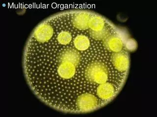
Chapter 9 Multicellular and tissue organization. Figure 9.1. Physalia physalis Portuguese Man-of-War. Figure 9.2. I. Intro - Origins of Multicellularity Multicellular life has been on earth for 550 million years, only ~10% of earth’s geological history

E N D
Figure 9.1 Physalia physalis Portuguese Man-of-War
I. Intro - Origins of Multicellularity • Multicellular life has been on earth for 550 million years, only ~10% of earth’s geological history • colonial hypothesis – multicellularity began as dividing cells remained together, as do many colonial protests (fig. 9.3) • syncytial hypothesis – multicellularity evolved from large, multinucleated cells that developed internal plasma membranes • Both of these happen in different protests
Are animals polyphyletic or monophyletic? The nearly simultaneous appearance of all animal phyla makes it hard to tell 1. if animals are polyphyletic, more than one explanation of the origins of multicellularity is possible 2. more than one body form could be ancestral 3. however, impressive similarities in animal cell organization support monophyletic origin (eg asters in cell division, cell junctions are similar in all animal cells, most animals produce flagellated sperm and most animal cells use similar proteins to accomplish movement)
Figure 9.3 Two hypotheses regarding the origin of Multicellularity
II. Phylum Porifera - Sponges • Primarily marine animals that consist of loosely organized cells; approx 9k spp, from < 1cm to > 1m
Figure 9.4 (a) Verongia
Figure 9.4 (b) Axiomella
A. Characteristics of members of Phylum Porifera include: 1. asymmetrical or radial symmetry 2. 3 types of cells - pinacocytes, mesenchyme cells (amoebocytes) and choanocytes 3. Central cavity or several branching chambers, thru which water flows for filter feeding 4. no tissues or organs
B. Cell types, Body wall, and Skeletons 1. sponge cells are specialized for particular functions (division of labor) • Pinacocytes • Mesenchyme • Choanocytes
a. pinacocytes - flat, thin cells that line the outer surface of a sponge. Pinacocytes may be slightly contractile and help sponge change shape. Some pinacocytes specialized into porocytes, which regulate water circulation (fig. 9.5) b. jelly like layer under pinacocytes is termed mesohyl. Mesenchyme cells are amoeboid, and move about in the mesohyl. Specialized for reproduction, transporting and storing food, secreting skeletal elements (spicules) c. beneath mesenchyme, lining inner chambers are choanocytes - collar cells. Flagellated cells with ring of microvilli surrounding flagella. Microfilaments connect microvilli, forming a net that helps filter edible particles (Fig. 9.5)
Figure 9.5 Morphology of a Simple Sponge
C. Sponges are supported by skeleton that may consist of spicules - needlelike spikes. • spicules are formed by amoeboid cells • made of CaCO3 or silica 3. may take on a variety of shapes ( fig. 9.6) 4. alternatively, skeleton may be made of spongin, a fibrous protein made of collagen - dried beaten and washed to produce commercial sponges
Figure 9.6 Sponge Spicules
D. Water currents and body forms - sponges lives depend on the water currents that choanocytes create 1. water brings food and O2, removes wastes • methods of food filtration and circulation reflect body forms in the phylum. 3 types: (fig. 9.7) • Ascon body form • Sycon body form • Leucon body form
i. ascon body form - simplest and least common. Vaselike form; • 1. ostia are outer openings of porocytes and lead directly to chamber called spongocoel • 2. choanocytes line spongocoel and their flagellar movements draw water into the spongocoel thru the ostia • 3. water exits sponge thru osculum, single large opening at the top of the sponge
ii. sycon body form - sponge wall appears folded • 1. water enters thru dermal pores, which are openings of incurrent canals • .pores in body walls open to radial canals, and radial canals lead to spongocoel • .choanocytes line radial canals and beating of flagella moves water from ostia, thru incurrent and radial canals, to spongocoel and out the osculum.
iii. leucon body forms have an extensively branched canal system. • 1. Water enters the ostium and moves thru branched incurrent canals, • 2. incurrent canals lead to choanocyte lined chambers. Canals leading away from the chambers are called excurrent canals3. proliferation of chambers and canals has resulted in absence of spongocoel. Often there are multiple exit points for water leaving sponge
Maintenance functions • 1, sponges feed on particles that range in size from .1 to 50 um. • a. bacteriab. microscopic algaec. protistsd. other suspended particles
2. important in reducing coastal turbidity • a. 1 leucon sponge, 1 cm in diameter and 10 cm high, filters 20 liters of water/day! 3. a few sponges are carnivorous - catch small crustaceans (deep water) with spicule-covered filaments .
4. feeding methods - choanocytes filter small suspended particles. • a. Water passes thru collar near base and moves into spongocoel at open end of collar • b. suspended food is trapped on collar and moved along microvilli to base of collar, where it is incorporated into a food vacuole • c. lysozymal enzymes and pH changes digest particle in vacuoled. partly digested food passed to amoeboid cells, that distribute it. 5. other feeding methods - • a. pinacocytes lining incurrent canals may phagocytize larger food particles. Sponges may also absorb nutrients in sea water thru active transport
Reproduction - most sponges are monoecious - both sexes occur in same individual; do not usually self fertilize because eggs and sperm ready at different times. • 1. certain choanocytes lose collars and flagella and undergo meiosis to form flagellated sperm2. other choanocytes may undergo meiosis and form eggs. Eggs retained in mesohyl of parent
3. sperm cells exit one sponge by osculum and enter another with incurrent water. they are trapped by choanocytes and put in vacuoles. • 4.sperm lose collar and flagella, become ameboid and transfer sperm to eggs • 5. early development occurs in mesohyl, then a flagellated larva forms. Larva breaks free, free-swims for up to 2 days before settling to substrate and develops into adult form (Fig. 9.8)
Figure 9.8 (abc) Flagellated cells cover outer surface Development of Sponge Larval Stages choanocytes pinacocytes
III. Phylum Cnidaria A. Intro - Members of Phylum Cnidaria possess radial symmetry -advantageous in sedentary animals because the sensory receptors are evenly distributed around the body - can respond to stimuli from all directions1. there are > 9k spp of Cnidarians, most are marine. Many important in coral reef ecosystems
2. Characteristics include: • a. radial symmetry • b. diploblastic, tissue level organization • c. gelatinous mesoglea between epidermal and gastrodermal tissue layers • d. gastrovascular cavity • e. nervous system in form of a netf. specialized cells called cnidocytes used in defense, feeding, and attachment
B. Body Wall and nematocysts • 1. diploblastic tissue organization - cells organize into tissues that can carry out more complex functions than individual cells; all cells derived from 2 embryological layers • 2. ectoderm of embryo gives rise to epidermis, endoderm gives rise to inner layer, called gastrodermis • a. cells differentiate into specialized cells for protections, food gathering, coordination, movement, digestion, and absorption
3. between the 2 layers is a jellylike layer called the mesoglea; cells present in this layer come from either epidermis or gastrodermis • 4. cnidocytes - cells characteristic of the phylum - Epidermal and gastrodermal cells both give rise to cnidocytes. Cnidocytes produce structures called nematocysts - feeding, defense, attachment.
5. a nematocyst is a fluid filled intracellular capsule enclosing a coiled, hollow tube (Fig. 9.10). A lid-like operculum covers capsule at one end. The cnidocyte has a modified cilium at the end, the trigger. If stimulated, it ejects the coiled tube within, like a sweater sleeve turned inside out. • a. cnidocysts may have spines to penetrate prey • b. some have toxins that are injected to paralyze prey • c. others have unarmed tubes that wrap around prey or substrate for attachmentd. some have sticky secretions to anchor itself. 6 or more types of nematocysts may be found on one individual
C. Alternation of Generations - most cnidarians possess 2 body forms in their life histories • 1. polyp - usually asexual and sessile; attaches to substrate at base, column (cylindrical body form) is capped by a mouth surrounded by tentacles • 2. medusa is dioecious and free swimming. shaped like inverted bowl, tentacles hang from rim. Mouth is centrally located facing downward, and medusa swims by pulsating body walls. More mesoglea in medusas than in polyps
Figure 9.11 Generalized Cnidarian Life Cycle
E. Reproduction - most are dioecious - each has a particular gender. • 1. sperm and eggs are released into gv cavity or to the outside. In some cases, eggs stay in mom till fertilization; embryo enlarges to form a ciliated, free swimming larva called a planula. Planula attaches to substrate, interior cells split to form gastrpvascular cavity and polyp develops • 2. medusae nearly always form from budding from body wall of polyp and polyps form other polyps by budding. buds may detach, or remain attached to contribute to a colony.
F. Class Hydrozoa - hydras - small relatively common cnidarians; most marine, but some are freshwater -characteristics: cnidocysts in epidermis; release sperm and eggs out of body • 1, most hydrozoans have alternation of generations, but in some, medusa stage is lost; in others, polyp stage is very small • 2. most hydrozoans are colonial w/ some individuals specialized for feeding and others specialized for defense or reproduction e.g Obelia3. Gonionemus has a mostly medusa form, living in shallow marine waters.4. Hydra is a freshwater hydrozoan that hangs from underside of floating plants in streams and ponds - lacks medusa stage
Figure 9.14 (a) G. Class Scyphozoa - true jellyfish - dominant life stage is medusa; cnidocytes in epidermis and gastrodermis layer a) Mastigias b) Aurelia
Figure 9.16 Aurelia life history (dioecious)
Class Cubozoa- Sea wasp Figure 9.17
Figure 9.18 (a) H. Class Anthozoa - anemones and corals - colonial or solitary, and lack medusae. Differ from hydrozoans bc sperm and eggs released into gastrovascular cavity and expelled from there
Figure 9.19 Class Anthozoa- structure of an Anemone
Figure 9.20 Class Anthozoa
Figure 9.21 (a) Other anthozoan- Octacorallian coral (Fleshy sea pen)
Figure 9.21 (b) Octacorallian Coral- (Purple sea fan)
Figure 9.22 (a) Phylum Ctenophora- Mnemiopsis known for the bioluminescence