Download
1 / 50
530 likes | 770 Views
CHAPTER 4 A Tour of the Cell. Modules 4.1 – 4.5. The Art of Looking at Cells. Artists are often inspired by biology and biology depends on art The paintings of Wassily Kandinsky (1866-1944) show the influence of cellular forms.

E N D
CHAPTER 4A Tour of the Cell Modules 4.1 – 4.5
The Art of Looking at Cells • Artists are often inspired by biology and biology depends on art • The paintings of Wassily Kandinsky (1866-1944) show the influence of cellular forms
Illustration is an important way to represent what scientists see through microscopes • The anatomist Santiago Ramón y Cajal (1852-1934) was trained as an artist • He drew these retina nerve cells
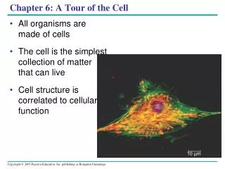
INTRODUCTION TO THE WORLD OF THE CELL • The microscope was invented in the 17th century • Using a microscope, Robert Hooke discovered cells in 1665 • All living things are made of cells (cell theory)
4.1 Microscopes provide windows to the world of the cell • The light microscope enables us to see the overall shape and structure of a cell Image seen by viewer Eyepiece Ocularlens Objective lens Specimen Condenser lens Light source Figure 4.1A
Electron microscopes were invented in the 1950s • They use a beam of electrons instead of light • The greater resolving power of electron microscopes • allows greater magnification • reveals cellular details
Scanning electron microscope (SEM) • Scanning electron micrograph of cilia Figure 4.1B
Transmission electron microscope (TEM) • Transmission electron micrograph of cilia Figure 4.1C
4.2 Cell sizes vary with their function • Below is a list of the most common units of length biologists use (metric) Table 4.2
Cell size and shape relate to function Figure 4.2
4.3 Natural laws limit cell size • At minimum, a cell must be large enough to house the parts it needs to survive and reproduce • The maximum size of a cell is limited by the amount of surface needed to obtain nutrients from the environment and dispose of wastes
A small cell has a greater ratio of surface area to volume than a large cell of the same shape 30 µm 10 µm Surface areaof one large cube= 5,400 µm2 Total surface areaof 27 small cubes= 16,200 µm2 Figure 4.3
4.4 Prokaryotic cells are small and structurally simple • There are two kinds of cells: prokaryotic and eukaryotic • Prokaryotic cells are small, relatively simple cells • They do not have a nucleus
A prokaryotic cell is enclosed by a plasma membrane and is usually encased in a rigid cell wall • The cell wall may be covered by a sticky capsule Prokaryoticflagella Ribosomes Capsule Cell wall • Inside the cell are its DNA and other parts Plasma membrane Nucleoid region(DNA) Pili Figure 4.4
4.5 Eukaryotic cells are partitioned into functional compartments • All other life forms are made up of one or more eukaryotic cells • These are larger and more complex than prokaryotic cells • Eukaryotes are distinguished by the presence of a true nucleus
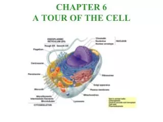
Smooth endoplasmicreticulum Nucleus Roughendoplasmicreticulum • An animal cell Flagellum Not in most plant cells Lysosome Centriole Ribosomes Peroxisome Golgiapparatus Microtubule Plasma membrane Cytoskeleton Intermediatefilament Microfilament Mitochondrion Figure 4.5A
The plasma membrane controls the cell’s contact with the environment • The cytoplasm contains organelles • Many organelles have membranes as boundaries • These compartmentalize the interior of the cell • This allows the cell to carry out a variety of activities simultaneously
A plant cell has some structures that an animal cell lacks: • Chloroplasts • A rigid cell wall
Roughendoplasmicreticulum Nucleus Ribosomes Smoothendoplasmicreticulum Golgiapparatus Microtubule Centralvacuole Not inanimalcells Intermediatefilament Cytoskeleton Chloroplast Microfilament Cell wall Mitochondrion Peroxisome Plasma membrane Figure 4.5B
ORGANELLES OF THE ENDOMEMBRANE SYSTEM 4.6 The nucleus is the cell’s genetic control center • The largest organelle is usually the nucleus • The nucleus is separated from the cytoplasm by the nuclear envelope • The nucleus is the cellular control center • It contains the DNA that directs the cell’s activities
NUCLEUS Chromatin Two membranesof nuclearenvelope Nucleolus Pore ROUGHENDOPLASMICRETICULUM Ribosomes Figure 4.6
4.7 Overview: Many cell organelles are related through the endomembrane system • The endomembrane system is a collection of membranous organelles • These organelles manufacture and distribute cell products • The endomembrane system divides the cell into compartments • Endoplasmic reticulum (ER) is part of the endomembrane system
Transport vesiclebuds off 4 Ribosome Secretory(glyco-) proteininside transportvesicle Sugarchain 3 Glycoprotein 1 2 ROUGH ER Polypeptide 4.8 Rough endoplasmic reticulum makes membrane and proteins • The rough ER manufactures membranes • Ribosomes on its surface produce proteins Figure 4.8
4.9 Smooth endoplasmic reticulum has a variety of functions • Smooth ER synthesizes lipids • In some cells, it regulates carbohydrate metabolism and breaks down toxins and drugs
SMOOTH ER ROUGHER Nuclearenvelope Ribosomes SMOOTH ER ROUGH ER Figure 4.9
4.10 The Golgi apparatus finishes, sorts, and ships cell products • The Golgi apparatus consists of stacks of membranous sacs • These receive and modify ER products, then send them on to other organelles or to the cell membrane
The Golgi apparatus Golgi apparatus Golgiapparatus “Receiving” side ofGolgi apparatus Transportvesiclefrom ER Newvesicleforming “Shipping”side of Golgiapparatus Transport vesiclefrom the Golgi Figure 4.10
4.11 Lysosomes digest the cell’s food and wastes • Lysosomes are sacs of digestive enzymes budded off the Golgi LYSOSOME Nucleus Figure 4.11A
digest food • destroy bacteria • recycle damaged organelles • function in embryonic development in animals • Lysosomal enzymes
Rough ER Transport vesicle(containing inactivehydrolytic enzymes) Plasmamembrane Golgiapparatus Engulfmentof particle Lysosomeengulfingdamagedorganelle “Food” LYSOSOMES Digestion Foodvacuole Figure 4.11B
4.12 Connection: Abnormal lysosomes can cause fatal diseases • Lysosomal storage diseases are hereditary • They interfere with other cellular functions • Examples: Pompe’s disease, Tay-Sachs disease
4.13 Vacuoles function in the general maintenance of the cell • Plant cells contain a large central vacuole • The vacuole has lysosomal and storage functions Centralvacuole Nucleus Figure 4.13A
Nucleus Contractilevacuoles • These pump out excess water • Protists may have contractile vacuoles Figure 4.13B
4.14 A review of the endomembrane system • The various organelles of the endomembrane system are interconnected structurally and functionally Transport vesiclefrom Golgi Transport vesiclefrom ER Rough ER Plasmamembrane Vacuole Nucleus Lysosome Golgiapparatus Smooth ER Nuclearenvelope Figure 4.14
ENERGY-CONVERTING ORGANELLES 4.15 Chloroplasts convert solar energy to chemical energy • Chloroplasts are found in plants and some protists • Chloroplasts convert solar energy to chemical energy in sugars Chloroplast Stroma Inner and outer membranes Granum Intermembranespace Figure 4.15
4.16 Mitochondria harvest chemical energy from food • Mitochondria carry out cellular respiration • This process uses the chemical energy in food to make ATP for cellular work
MITOCHONDRION Outermembrane Intermembranespace Innermembrane Cristae Matrix Figure 4.16
THE CYTOSKELETON AND RELATED STRUCTURES 4.17 The cell’s internal skeleton helps organize its structure and activities • A network of protein fibers makes up the cytoskeleton Figure 4.17A
Intermediate filaments reinforce the cell and anchor certain organelles • Microtubules • give the cell rigidity • provide anchors for organelles • act as tracks for organelle movement • Microfilaments of actin enable cells to change shape and move
Tubulinsubunit Actin subunit Fibrous subunits 25 nm 7 nm 10 nm MICROFILAMENT INTERMEDIATEFILAMENT MICROTUBULE Figure 4.17B
4.18 Cilia and flagella move when microtubules bend • Eukaryotic cilia and flagella are locomotor appendages that protrude from certain cells • A cilia or flagellum is composed of a core of microtubules wrapped in an extension of the plasma membrane
FLAGELLUM Electron micrograph of sections: Outer microtubule doublet Plasmamembrane Flagellum Centralmicrotubules Outer microtubule doublet Plasmamembrane Basal body Basal body(structurally identical to centriole) Figure 4.18A
Microtubule doublet • Clusters of microtubules drive the whipping action of these organelles Slidingforce Dynein arm Figure 4.18B
EUKARYOTIC CELL SURFACES AND JUNCTIONS 4.19 Cell surfaces protect, support, and join cells • Cells interact with their environments and each other via their surfaces • Plant cells are supported by rigid cell walls made largely of cellulose • They connect by plasmodesmata, channels that allow them to share water, food, and chemical messages
Walls of two adjacent plant cells Vacuole PLASMODESMATA Layers of one plant cell wall Cytoplasm Plasma membrane Figure 4.19A
Animal cells are embedded in an extracellular matrix • It is a sticky layer of glycoproteins • It binds cells together in tissues • It can also have protective and supportive functions
Anchoring junctions link animal cells • Communicating junctions allow substances to flow from cell to cell • Tight junctions can bind cells together into leakproof sheets TIGHTJUNCTION ANCHORING JUNCTION COMMUNICATING JUNCTION Plasma membranes ofadjacent cells Extracellularmatrix Figure 4.19B
4.20 Eukaryotic organelles comprise four functional categories • Eukaryotic organelles fall into four functional groups Table 4.20
4.21 Connection: Extraterrestrial life-forms may share features with life on Earth • It is almost certain that Earth is the only life-bearing planet in our solar system • But it is conceivable that conditions on some of the moons of the outer planets or on planets in other solar systems have allowed the evolution of life Figure 4.21