Download
1 / 32
360 likes | 5.48k Views
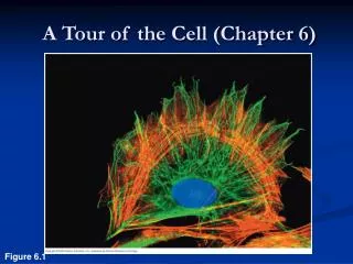
CHAPTER 7 A TOUR OF THE CELL. Section A: How We Study Cells 1. Microscopes provide windows to the world of the cell Cell biologists can isolate organelles to study their function. Objectives. Distinguish between prokaryotic and eukaryotic cells

E N D
CHAPTER 7A TOUR OF THE CELL Section A: How We Study Cells 1. Microscopes provide windows to the world of the cell Cell biologists can isolate organelles to study their function
Objectives • Distinguish between prokaryotic and eukaryotic cells • Explain why there are upper and lower limits to cell size • Explain the function of compartmentalization in eukaryotic cells
Describe the structure and function of the nucleus • Describe the structure and function of the eukaryotic ribosome • List the components of the endomembrane system, describe their structures and functions and summarize the relationships among them • Describe the types of vacuoles and how their functions differ
Explain the role of peroxisomes in eukaryotic cells • Describe the structure of a typical mitochondrion, detail its function and explain how compartmentaliztion in the mitochondrion is important to this function • Explain the structure and functioning of the chloroplast
Describe the functions of the cytoskeleton and distinguish among microtubules, microfilaments and intermediate filaments • Describe the structure of flagella and cilia and briefly summarize the relationship between this structure and their functioning
1. Microscopes provide windows to the world of the cell • The discovery and early study of cells progressed with the invention and improvement of microscopes in the 17th century. • In a light microscope (LMs) visible lightpasses through the specimen and then through glass lenses. • The lenses refract light such that the image is magnified into the eye or a video screen. • A light microscope can be used to resolve individual cells
Microscopes vary in magnification and resolving power. • Magnification is the ratio of an object’s image to its real size. • Resolving power is a measure of image clarity. • It is the minimum distance two points can be separated and still viewed as two separate points. • Resolution is limited by the shortest wavelength of the source, in this case light.
The minimum resolution of a light microscope is about 2 microns, the size of a small bacterium • Light microscopes can magnify effectively to about 1,000 times the size of the actual specimen. • At higher magnifications, the image blurs. Fig. 7.1
A light microscope can resolve individual cells but it cannot resolve much of the internal anatomy, especially the organelles. • To resolve smaller structures we use an electron microscope (EM), which focuses a beam of electrons through the specimen or onto its surface. • the practical limit of a modern EM is about about 2 nm (the size of a single rhinovirus).
Transmission electron microscopes (TEM) are used mainly to study the internal ultrastructure of cells. • A TEM aims an electron beam through a thin section of the specimen. • The image is focused and magnified by electromagnets. • To enhance contrast, the thin sections are stained with atoms of heavy metals. Fig. 7.2a
Scanning electron microscopes (SEM) are useful for studying surface structures. • The sample surface is covered with a thin film of gold. • The beam excites electrons on the surface. • These secondary electrons are collected and focused on a screen. • The SEM has great depth of field, resulting in an image that seems three-dimensional. Fig. 7.2b
Electron microscopes reveal organelles, but they can only be used on dead cells and they may introduce some artifacts. • Light microscopes do not have as high a resolution, but they can be used to study live cells. • Microscopes are a major tool in cytology, the study of cell structures. • Cytology + biochemistry = modern cell biology.
Cell Theory • All known living things are made up of cells. • The cell is structural & functional unit of all living things. • All cells come from pre-existing cells by division. (Spontaneous Generation does not occur). 1838: Schleiden and Schwann proposed cell theory These first three are the very basic foundations of Cell Theory. All six of these components make up modern Cell Theory • Cells contains hereditary information which is passed from cell to cell during cell division. • All cells are basically the same in chemical composition. • All energy flow (metabolism & biochemistry) of life occurs within cells.
2. Cell biologists can isolate organelles to study their functions • The goal of cell fractionation is to separate the major organelles of the cells so that their individual functions can be studied.
Uses an ultracentrifuge, a machine that can spin at up to 130,000 revolutions per minute and apply forces more than 1 million times gravity (1,000,000 g). • Fractionation begins with homogenization, gently disrupting the cell. • Then, the mixture is spun in a centrifuge to separate heavier pieces into the pellet while lighter particles remain in the solution. • As the process is repeated at higher speeds and longer durations, smaller and smaller organelles can be collected in subsequent pellets.
Cell fractionation prepares quantities of specific cell components. • The functions of these organelles to be isolated, especially by the reactions or processes catalyzed by their proteins. • For example, one cellular fraction is enriched in enzymes that function in cellular respiration. • Electron microscopy reveals that this fraction is rich in the organelles called mitochondria. • Cytology and biochemistry complement each other in connecting cellular structure and function.
Section B: A Panoramic View of the Cell 1. Prokaryotic and eukaryotic cells differ in size and complexity 2. Internal membranes compartmentalize the functions of a eukaryotic cell
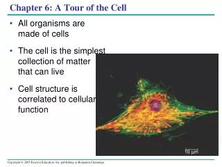
1. Prokaryotic and eukaryotic cells differ in size and complexity • All cells are surrounded by a plasma membrane. (What is this made of?) • The “liquid” inside the membrane is the cytosol, which contains the organelles. • All cells contain chromosomes which have genes in the form of DNA. • All cells also have ribosomes, organelles that make proteins using the instructions contained in genes.
A major difference between prokaryotic and eukaryotic cells is the location of chromosomes. • In an eukaryotic cell, chromosomes are contained in a membrane-enclosed organelle, the nucleus. • In a prokaryotic cell, the DNA is concentrated in the nucleoid without a membrane separating it from the rest of the cell.
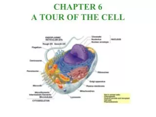
Fig. 7.4 The prokaryotic cell is much simpler in structure, lacking a nucleus and the other membrane-enclosed organelles of the eukaryotic cell.
CD-Rom Activity 7.1 • This activity will help you to review and gain an understanding of the structures and functions of prokaryotic cells.
In eukaryote cells, the chromosomes are contained within a membranous nuclear envelope. • The region between the nucleus and the plasma membrane is the cytoplasm. (Sarah, this is for you.) • All the material within the plasma membrane of a prokaryotic cell is cytoplasm. (This includes the organelles.) • Within the cytoplasm of a eukaryotic cell is a variety of membrane-bounded organelles of specialized form and function. • These membrane-bounded organelles are absent in prokaryotes.
Eukaryotic cells are generally much bigger than prokaryotic cells. • The logistics of carrying out metabolism set limits on cell size. • At the lower limit, the smallest bacteria, mycoplasmas, are between 0.1 to 1.0 micron. • Most bacteria are 1-10 microns in diameter. • Eukaryotic cells are typically 10-100 microns in diameter.
Metabolic requirements also set an upper limit to the size of a single cell. • As a cell increases in size its volume increases faster than its surface area. • Smaller objects have a greater ratio of surface area to volume. Fig. 7.5
The plasma membrane functions as a selective barrier that allows passage of oxygen, nutrients, and wastes for the whole volume of the cell. Fig. 7.6
The volume of cytoplasm determines the need for this exchange. • Rates of chemical exchange may be inadequate to maintain a cell with a very large cytoplasm. • The need for a large surface to accommodate the volume explains the microscopic size of most cells. • Larger organisms do not generally have larger cells than smaller organisms - simply more cells.
2. Internal membranes compartmentalize the functions of a eukaryotic cell • A eukaryotic cell has extensive and elaborate internal membranes, which partition the cell into compartments. • These membranes also participate in metabolism as many enzymes are built into membranes. • The barriers created by membranes provide different local environments that facilitate specific metabolic functions.
The general structure of a biological membrane is a double layerof phospholipids with other lipids and diverse proteins. • Each type of membrane has a unique combination of lipids and proteins for its specific functions. • For example, those in the membranes of mitochondria function in cellular respiration.
CD-Rom Activity 7.2 • This activity will help you to review and gain an understanding of the structures and functions of animal cells.
CD-Rom Activity 7.3 • This activity will help you to review and gain an understanding of the structures and functions of plant cells.