Download
1 / 14
170 likes | 380 Views
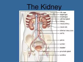
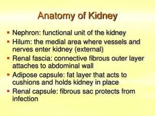
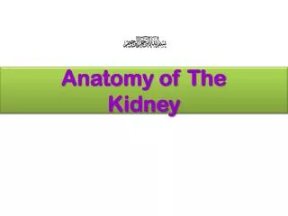
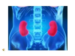
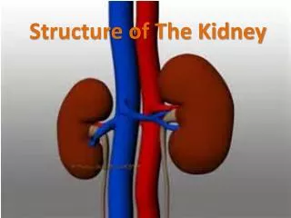
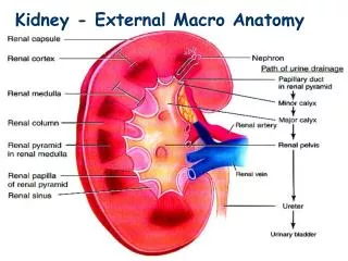
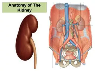
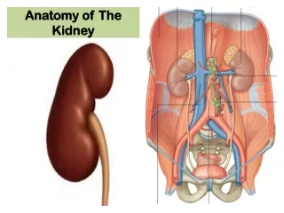
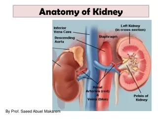
Renal Cortex Outer layer Renal Medulla Cone shaped tissue called renal pyramids Renal Pelvis Calyxes cup-shaped tubes Central cavity that is continuous with ureter. Anatomy of the Kidney. Kidney is composed of millions of NEPHRONS Functional unit of kidney

E N D
Renal Cortex • Outer layer • Renal Medulla • Cone shaped tissue called renal pyramids • Renal Pelvis • Calyxes cup-shaped tubes • Central cavity that is continuous with ureter Anatomy of the Kidney
Kidney is composed of millions of NEPHRONS • Functional unit of kidney • Each composed of a system of tubules and has its own blood supply Anatomy of a Nephron
Afferent arteriole • Takes blood (via the renal artery) to the glomerulus • Glomerulus • Knot of capillaries in side Bowman’s Capusule • Made up of podocyte cells that allow small molecules to be filtered through Parts of a nephron
Efferent arteriole • Transports filtered blood to the capillary network that surrounds the nephron • Tubules • Proximal Convoluted Tubule (PCT) • Tubular reabsorption occurs • Loop of Henle • Distal Convoluted Tubule (DCT) • Tubular secretion occurs Parts of a nephron
Collecting Duct • From the DCT the filtrate enters the collecting duct where it is taken to the renal pelvis Parts of a nephron
1. Glomerular Filtration • Blood enters the afferent arteriole into the glomerulus • Here water and small molecules are filtered into Bowman’s capsule • Water, nutrients, salts, waste molecules are filtered and called the filtrate • Large molecules like blood cells and platelets can’t pass through and exit vis efferent arteriole Urine formation
Body filters approx. 180 L of water/day • If the composition of urine were the exact same as the filtrate then our body would continually lose a large amount of water, salt and nutrients every time we went to the washroom! • This means our body must reabsorb nutrients and water is back into the body somewhere before we urinate Urine formation
2. Tubular Reabsorption • From Bowman’s Capsule the filtrate enters proximal convoluted tubule • Here molecules from the filtrate are reabsorbed back into the blood of the capillary network Urine formation
3. Tubular Secretion • From the PCT the filtrate enters the Loop of Henle, and then finally into the distal convoluted tubule where secretion occurs • Here wastes from the blood that were not filtered through Bowman’s capsule enter the tubule • Ammonia and many drugs are removed from the blood during secretion • From here it travels to the collecting duct where it is transported out of the body via the bladder Urine formation
Filtration • Blood is filtered • Filtrate enters the nephron tubules • Wastes enter blood • Reabsorption • Molecules from filtrate are reabsorbed back into blood (body needs them to function) • Secretion • Wastes in the blood secreted back into the nephron for removal Urine Formation