Download

1 / 25
580 likes | 2.54k Views
Anatomy of The Kidney & Ureter. Dr. Nimir Dr. Safaa. Objectives. Describe the normal site, size, shape & position of the kidney. Identify the borders, poles and surfaces of the kidney. Delineate the surface anatomy of the kidney.

E N D
Anatomy of The Kidney & Ureter Dr. Nimir Dr. Safaa
Objectives • Describe the normal site, size, shape & position of the kidney. • Identify the borders, poles and surfaces of the kidney. • Delineate the surface anatomy of the kidney. • Discuss the structures passing through the hilum of the kidney in order. • Discuss the peritoneal covering and relations of the two kidneys. • Describe the fascia surrounding the kidney. • Discuss the blood and nerve supply of the kidney. • Describe the kidney segmentation. • Discuss the lymphatic drainage of the kidney.
Objectives contd. • Describe the beginning, termination and parts of the ureter. • Compare between the course and relations of both abdominal & pelvic ureter. • Delineate the surface anatomy of abdominal ureter. • Discuss blood and nerve supply, and lymphatic drainage of ureter. • Discuss the normal sites of the ureteric constrictions.
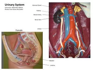
The urinary system consists of the following • Kidneys • Ureters • Urinary bladder • Urethra
The Kidney • Bean-shaped organ • It lies on the posterior abdominal wall at the side of vertebral column • It measures 4x2x1 inches • It has anterior and posterior surfaces and medial and lateral borders • Right kidney is lower than the left because of the liver • The level of kidneys varies with respiration of about 1 inch
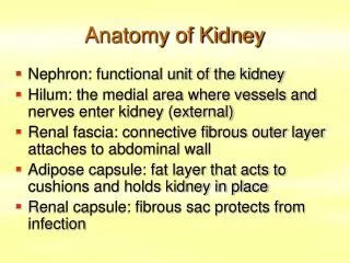
The Kidney Cont., Renal hilum • Is found at the medial concave surface • Renal vein, renal artery and ureter are found in the hilum in anterior posterior order • Lymphatics and sympathetic nerves pass through the hilum Renal sinus All structures pass through the hilum in addition to small amount of fat and the pelvis are found in the sinus
Suprarenal glands, kidneys, and ureters and their vessels are retroperitoneal structures • Renal capsule formed of dense connective tissue surrounding the kidneys for support and protection • Perirenal fat a layer of fat surrounds the kidney outside the renal capsule
Kidneys, suprarenal glands, and perirenal fat are enclosed by fascial membrane called Renal fascia • The two layers extend medially to enclose the renal vessels and blend with vascular fascia • The two layers extend inferiorly to enclose the ureters as asPeriuretric fascia • A layer of fat surrounding the kidneys, ureters, suprarenal glands external to renal fascia called Pararenal fat. • Perirenal fat, renal fascia, pararenal fat and some collagen fibers hold the kidneys in fixed position • Superiorly, renal fascia is attached to inferior diaphragmatic fascia
Internal structure of the Kidney Each kidney consists of: • Outer cortex • Inner medulla formed of renal pyramids with apex forming renal papilla • Cortex extends into medulla as renal columns • Renal pelvis fills most of the sinus • Major calyces • Minor calyces
Anterior relation of the Kidney Right kidney: • Suprarenal gland • Liver • Duodenum • Right colic flexure • Ileum Left kidney • Suprarenal gland • Stomach • Spleen • Splenic artery • Pancreas • Jejunum • Left colic flexure
Anterior relation of the Kidney Left Kidney Right Kidney
Posterior Relation of the Kidney • Left Kidney: • Diaphragm separates the kidney from the pleural cavity and 11th and 12th ribs • Psoas major medially • Quadratuslumborum in the middle, transversusabdominis laterally • Subcostal, iliohypogastric and ilioinguinal nerves descend obliquely behind the kidney
Posterior Relation of the Kidney • Right Kidney: • Diaphragm separates the kidney from the pleural cavity and 12th rib. • Psoas major medially • Quadratuslumborum in the middle, transversusabdominis laterally • Subcostal, iliohypogastric and ilioinguinal nerves descend obliquely behind the kidney
Blood Supply of the Kidney • Renal arteries are paired branches arise from aorta at the level of intervertebral disc between L1,2 • Right one longer than the left, passes behind IVC • At the hilum, each one divides into 5 segmental arteries each supplies a renal segment • Renal segments are independent in their blood supply • Blood is drained by segmental veins to renal veins
Lymph Drainage • Lymph drains to the lateral aortic lymph nodes around the origin of the renal artery. • Nerve Supply • The nerve supply is the renal sympathetic plexus. The afferent fibers that travel through the renal plexus enter the spinal cord in the 10th, 11th, and 12th thoracic nerves.
The Ureter • It is a muscular organ • Extends from renal pelvis in abdomen, crosses the pelvic brim at common iliac artery bifurcation to the urinary bladder • It 10 inches in length • On the back, extends along a line from a point 5 cm from spine of L1 to posterior superior iliac spine
The Ureter Cont., • The abdominal part is retroperitoneal • It runs close to the tips of transverse processes of lumbar vertebrae as seen in contrast radiographs • It passes vertically on psoas muscle • It enters the pelvis crossing the external iliac artery • It shows three constriction along its course: Between ureter and pelvis At crossing the external iliac artery At entrance to urinary bladder • They are the sites of obstruction by renal calculi
The Ureter Cont., • Ureter runs on the lateral part of lesser pelvis • It runs parallel to medial part of greater sciatic notch • At level of ischial spine, it turns anteromedially to enter the inferior surface of the bladder in inferiomedial direction • The entrance is 5 cm a part on external surface and 2.5 cm a part at internal surface • Oblique passage creates a sphincter like structure at lower end of ureter
Anterior Relation of the Ureters • Right Ureter • The duodenum • The terminal part of the ileum • The right colic and ileocolic vessels • The right testicular or ovarian vessels(gonadal) • The root of the mesentery of the small intestine • Left Ureter • The sigmoid colon and sigmoid mesocolon • the left colic vessels • the left testicular or ovarian vessels
Anterior Relation of the Ureter Duodenum Terminal Ileum Left gonadal vessels Right gonadal vessels Left colic vessels Right colic vessels Sigmoid colon and mesocolon Ileocolic vessels
Posterior Relation of the Ureters • Right Ureter • The right psoas muscle, which separates it from the lumbar transverse processes • The bifurcation of the right common iliac artery • Left Ureter • The left psoas muscle, which separates it from the lumbar transverse processes • The bifurcation of the left common iliac artery
Blood Supply • Arteries • The arterial supply to the ureter is as follows: upper end, the renal artery; middle portion, the gonadal (testicular or ovarian) artery; and in the pelvis, the superior vesical artery. • Veins • Venous blood drains into veins that correspond to the arteries.
Lymph Drainage • The lymph drains to the lateral aortic nodes and the iliac nodes. • Nerve Supply • The nerve supply is the renal, testicular (or ovarian), and hypogastric plexuses (in the pelvis). Afferent fibers travel with the sympathetic nerves and enter the spinal cord in the first and second lumbar segments.