Download
1 / 21
210 likes | 223 Views
Gastrointestinal tract. Fine structure of the hollow and parenchymal viscera. Dr. Altdorfer. Splanchnology - the study of the visceral organs : digestive, r espiratory , u r ogenital systems. Parenchymal organs. Tubular (hollow) organs. Parenchymal organs. C.t. capsule

E N D
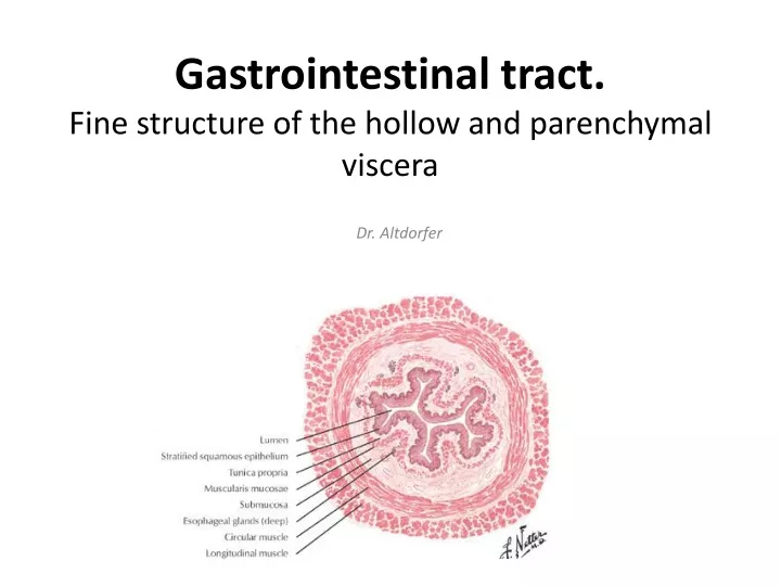
Gastrointestinal tract. Fine structure of the hollow and parenchymal viscera Dr. Altdorfer
Splanchnology - the study of the visceral organs: • digestive, • respiratory, • urogenital systems.
Parenchymal organs Tubular (hollow) organs
Parenchymalorgans C.t. capsule • Stroma / interstitium: c. t. network (reticular cells) • Parenchyme: e.g. glandular epithelium thymus lobule Liver (Azan) Interlobular septum lobule
Reticular cells Reticular fibres – AgNO3 impr.
Tubular organs – typical wall structure 1.Tunica mucosa
Tubular organs – wall structure 1.Tunica mucosa 2. Tela submucosa
Tubular organs – wall structure 1.Tunica mucosa 2. Tela submucosa 3. Tunica muscularis Stratumcirculare Stratumlongitudinale
Tubular organs – wall structure 1.Tunica mucosa 2. Tela submucosa 3. Tunica muscularis Stratumcirculare Stratumlongitudinale 4. Tela subserosa 5. Tunica serosa (mesothelium) or 4. Tunica adventitia
Tubular organs – wall structure 1.Tunica mucosa Epitheliummucosae Laminapropriamucosae Lamina muscularis mucosae (onlyin GI tract!) 2. Tela submucosa 3. Tunica muscularis Stratumcirculare Stratumlongitudinale 4. Tela subserosa 5. Tunica serosa (mesothelium) or 4. Tunica adventitia
a) Epithelium mucosae Str. basale (germinativum) Str. spinosum (polygonale) Str. superficiale (planocellulare) b) Lamina propria mucosae (Schaffer-glands) c) Lamina muscularismucosae(onlyin GI tract) a) Oesophagus1. Tunicamucosa b) c)
1. 3. Lamina propria mucosae 2. 2. 1. 1. • Lymphocyte • Plasma cells • Fibroblast, fibrocyte • Smooth muscle cells 4.
a) Epithelium mucosae Str. basale (germinativum) Str. spinosum (polygonale) Str. superficiale (planocellulare) b) Lamina propria mucosae (Schaffer-glands) c) Lamina muscularis mucosae a) esophagus1. Tunicamucosa b) c) 2. Tela submucosa (oesophagealglands) Submucous plexus of Meissner
Submucous plexus of Meissner
Stratum circulare • Stratum longitudinale 1/3 1/3 1/3 esophagus – 3. Tunicamuscularis Myenteric plexus of Auerbach
str. circulare str. longitudinale Tunicamuscularis Myenteric plexus of Auerbach
Tela subserosa, Tunica serosa (or: Adventitia )
Dr. Puskár Röhlich Pál: Szövettan, Budapest, 2006 Anthony L. Mescher: Junqueira’s Basic Histology, New York, 2010 Michael Ross and Lynn J. Romrell: Histology, Baltimore, 1989