Download
1 / 55
580 likes | 698 Views
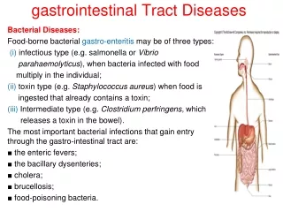
GASTROINTESTINAL TRACT. ESOPHAGUS. Clinical manifestations: 1-Dysphagia (difficulty in swallowing), which is attributed either to deranged esophageal motor function or to narrowing or obstruction of the lumen .

E N D
ESOPHAGUS • Clinical manifestations: • 1-Dysphagia (difficulty in swallowing), which is attributed either to deranged esophageal motor function or to narrowing or obstruction of the lumen. • 2-Heartburn (retrosternal burning pain) usually reflects regurgitation of gastric contents into the lower esophagus. • 3-Hematemesis (vomiting of blood) • 4-Melena (blood in the stools) are evidence of severe inflammation, ulceration, or laceration of the esophageal mucosa.
Achalasia • It means “failure to relax” • It is incomplete relaxation of the lower esophageal sphincter in response to swallowing. • This produces functional obstruction of the esophagus, with consequent dilation of the more proximal esophagus.
Manometric studies show 3 major abnormalities in achalasia: • (1) aperistalsis • (2) partial or incomplete relaxation of the lower esophageal sphincter with swallowing • (3) increased resting tone of the lower esophageal sphincter
Types of achalasia • 1-Primary achalasia • there is loss of intrinsic inhibitory innervation of the lower esophageal sphincter and smooth muscle segment of the esophageal body. • 2-Secondary achalasia • arise from pathologic processes that impair esophageal function. • 1-Chagas disease, caused by Trypanosoma cruzi, which causes destruction of the myenteric plexus of the esophagus, duodenum, colon, and ureter. • 2-Disorders of the dorsal motor nuclei such as polio, and autonomic neuropathy in diabetes
Primary achalasia • Progressive dilation of the esophagus above the level of the lower esophageal sphincter. • The wall of the esophagus may be of normal thickness, thicker than normal because of hypertrophy of the muscularis or markedly thinned by dilation. • The myenteric ganglia are usually absent from the body of the esophagus but may or may not be reduced in number in the region of the lower esophageal sphincter. • Inflammation in the location of the esophageal myenteric plexus is pathognomonic of the disease..
Clinical manifestation • stasis of food may produce mucosal inflammation and ulceration proximal to the lower esophageal sphincter. • progressive dysphagia and inability to completely convey food to the stomach. • Nocturnal regurgitation and aspiration of undigested food.
It usually becomes manifest in young adulthood, but it may appear in infancy or childhood. • The most serious outcome is esophageal squamous cell carcinoma (in about 5% of patients) and typically at an earlier age than in those without achalasia
Hiatal Hernia • separation of the diaphragmatic crura and widening of the space between the muscular crura and the esophageal wall permits a dilated segment of the stomach to protrude above the diaphragm.
Two anatomic patterns are recognized : • 1-the axial, or sliding, hernia (95%) . • 2-the nonaxial or paraesophageal, hernia.
In the sliding hernia protrusion of the stomach above the diaphragm creates a bell-shaped dilation, bounded below by the diaphragmatic narrowing.
In paraesophageal hernias, a separate portion of the stomach, usually along the greater curvature, enters the thorax through the widened foramen. • The cause of this deranged anatomy whether congenital or acquired is unknown.
hiatal hernias are reported in 1-20% of adult subjects, increasing in incidence with age. • ~ 9% of these adults suffer from heartburn or regurgitation of gastric juices into the mouth. • symptoms more likely result from incompetence of the lower esophageal sphincter than from the hiatal hernia per se. • symptoms accentuated by positions favoring reflux (bending forward, lying supine) and obesity.
Complications • 1-individuals with severe reflux esophagitis are likely to have a sliding hiatal hernia although most individuals with sliding hiatal hernias do not have reflux esophagitis. • 2- mucosal ulceration. • 3- bleeding. • 4- perforation.
5-Paraesophageal hernias can become strangulated or obstructed.
Lacerations (Mallory-Weiss Syndrome) • Longitudinal tears in the esophagus at the esophagogastric junction. • They are seen in : • 1-chronic alcoholics after a bout of severe retching or vomiting. • 2-during acute illnesses with severe vomiting.
Clinical manifestation • Esophageal lacerations account for 5-10% of upper GI bleeding episodes. • Most often bleeding is not profuse and ceases without surgical intervention, but life-threatening hematemesis may occur. • Healing is usually prompt with minimal to no residual problems.
Pathogenesis • inadequate relaxation of the musculature of the lower esophageal sphincter during vomiting with stretching and tearing of the esophagogastric junction at the moment of propulsive expulsion of gastric contents.
a hiatal hernia is found in more than 75% of patients with Mallory-Weiss tears. • almost half of individuals presenting with upper GI bleeding attributable to a Mallory-Weiss tear have no antecedent history of nausea, retching, abdominal pain, or vomiting. • normal variability in intra-abdominal pressure can be transduced through a hiatal hernia occasionally leading to a Mallory-Weiss tear.
Complications : • 1-Tears may involve only the mucosa or may penetrate the wall. • 2-Infection of the defect may lead to an inflammatory ulcer or to mediastinitis.
VARICES • One of the few potential sites for communication between the intra-abdominal splanchnic circulation and the systemic venous circulation is through the esophagus. • When portal venous blood flow into the liver is impeded by cirrhosis or other causes, the resultant portal hypertension induces the formation of collateral bypass channels wherever the portal and systemic systems communicate. • Portal blood flow is diverted through the stomach veins into the plexus of esophageal subepithelial and submucosal veins then into the azygos veins and the superior vena cava.
The increased pressure in the esophageal plexus produces dilated tortuous vessels called varices. • Persons with cirrhosis develop varices at a rate of 5-15 %/ year. • Varices are present in approximately 2/3 of all cirrhotic patients.
Variceal rupture produces: • 1- massive hemorrhage • 2- suffusion of blood into the esophageal wall
The conditions leading to initial rupture of a varix are unclear: • 1-silent erosion of overlying thinned mucosa • 2-increased tension in progressively dilated veins • 3-vomiting with increased intra-abdominal pressure
50% of those affected are found to have coexistent hepatocellular carcinoma. • Variceal hemorrhage subsides spontaneously in only 50% of cases. • 20-30% of patients die during the first episode of bleeding • Among those who survive, rebleeding occurs in approximately 70% within 1 year, with a similar rate of mortality for each episode.
ESOPHAGITIS • The inflammation may have many origins: • 1- reflux of gastric contents (reflux esophagitis). • 2- prolonged gastric intubation • 3--uremia • 4- ingestion of corrosive or irritant substances • 5- radiation • 6- chemotherapy
Reflux Esophagitis • Contributory factors for gastroesophageal reflux disease : • 1- Decreased efficacy of esophageal antireflux • mechanisms. • 2- CNS depressants • 3- Alcohol • 4- Tobacco • 5- Inadequate or slowed esophageal clearance of refluxed material • 6- The presence of a sliding hiatal hernia • 7- Increased gastric volume contributing to the volume of refluxed material • 8- Impaired reparative capacity of the esophageal mucosa by prolonged exposure to gastric juices
Incidence • In northern Iran the prevalence of esophagitis is more than 80% • It is also extremely high in regions of China. • Gastroesophageal reflux disease affects ~ 0.5% of the US adult population and has recurrent heartburn as its dominant symptom.
Clinical Features • Adults older than age 40 and occasionally infants and children • Heartburn • Regurgitation of a sour brash. • Attacks of severe chest pain mimicking a heart attack. • The severity of symptoms is not closely related to the presence and degree of anatomic esophagitis.
Complications of reflux esophagitis • 1- Bleeding • 2- Stricture formation • 3- Barrett esophagus with its predisposition to malignancy
Reflux esophagitis Numerous eosinophils (arrows) are present within the mucosa, and the stratified squamous epithelium has not undergone complete maturation because of ongoing inflammatory damage.
BARRETT ESOPHAGUS • Replacement of the normal distal stratified squamous mucosa by metaplastic columnar epithelium containing goblet cells. • It is a complication of long-standing gastroesophageal reflux. • Occurs in 5-15% of persons with persistent symptomatic or asymptomatic reflux disease. • It is unclear why individuals with few symptoms and little inflammation develop Barrett esophagus, and, conversely, why others have erosive esophagitis without Barrett esophagus
M:F ratio 4:1 • Common in whites more than in other races.
Pathogenesis • Prolonged and recurrent gastroesophageal reflux produce inflammation and eventually ulceration of the squamous epithelial lining. • Healing occurs by ingrowth of progenitor cells and re-epithelialization.
In the microenvironment of an abnormally low pH in the distal esophagus caused by acid reflux the cells differentiate into columnar epithelium. • Metaplastic columnar epithelium is thought to be more resistant to injury from refluxing gastric contents. • The metaplastic epithelium is not a typical intestinal epithelium as absorptive enterocytes.
Complications • 1-Ulcer • 2-Stricture • 3-Development of adenocarcinoma • Persons with Barrett esophagus have a 30-100X greater risk of developing esophageal adenocarcinoma than do normal populations, the greatest risk being associated with high-grade dysplasia.
A-Normal gastroesophageal junction B-The granular zone of Barrett esophagus (arrow)
Barrett esophagus Endoscopic view showing red velvety gastrointestinal-type mucosa extending from the gastroesophageal orifice.
Barrett esophagussquamous mucosa (left) and intestinal-type columnar epithelial cells in glandular mucosa (right).
ESOPHAGEAL CARCINOMA • Types: • 1-squamous cell carcinomas (90%) • 2-adenocarcinomas
In the USA 3-5X increase in the last 40 years in the incidence of adenocarcinomas associated with Barrett esophagus. • Adenocarcinoma arising in Barrett esophagus is more common in whites than in blacks. • Squamous cell carcinomas are more common in blacks worldwide.
In USA 6 new cases/100,000 population/year • In high incident areas as northern China and Iran, the prevalence is well over 100/ 100,000/year
Risk Factors for Squamous Cell Carcinoma of the Esophagus • 1-Esophageal Disorders • Long-standing esophagitis • Achalasia • Plummer-Vinson syndrome (esophageal webs, microcytic hypochromic anemia, atrophic glossitis)
2-Life-style • 1-prolong mucosal exposure to potential carcinogens such as those contained in tobacco and alcoholic beverages . • 2-chronic esophagitis which is often the consequence of alcohol and tobacco use.
Alcohol and tobacco use have well-defined predisposing role for chronic esophagitis. • These 2 agents are associated with the majority of squamous cell carcinoma in Europe and the United States.
3-Dietary • Deficiency of vitamins (A, C, riboflavin, thiamine, pyridoxine) • Deficiency of trace metals (zinc, molybdenum) • Fungal contamination of foodstuffs • High content of nitrites/nitrosamines
Diet, must underlie the very high incidence of esophageal SCC among the Moslems of Iran who neither drink nor smoke
The high levels of nitrosamines and fungi contained in some foods probably account for the very high incidence of this tumor in some regions of China. • A strong association with HPV occurs only in high-incidence areas.
4-Genetic Predisposition • Abnormalities affecting the p16/INK4 and EGFR are frequently present in squamous cell carcinoma of the esophagus.