Download
1 / 61
630 likes | 641 Views
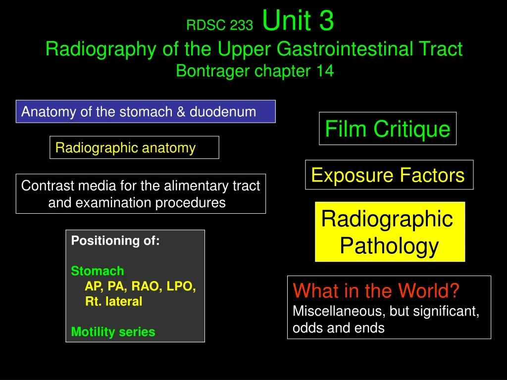
RDSC 233 Unit 3 Radiography of the Upper Gastrointestinal Tract Bontrager chapter 14. Anatomy of the stomach & duodenum. Film Critique. Radiographic anatomy. Exposure Factors. Contrast media for the alimentary tract and examination procedures.

E N D
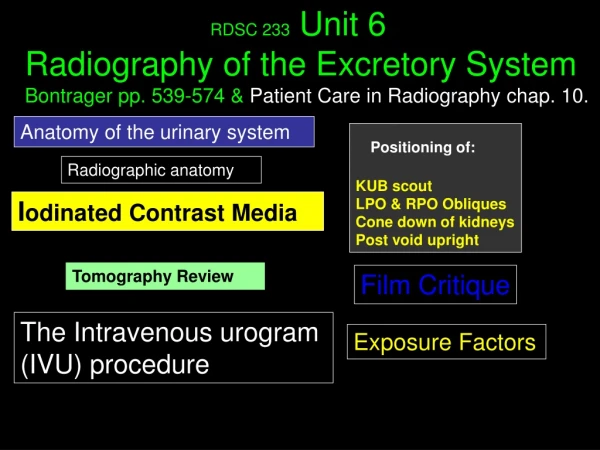
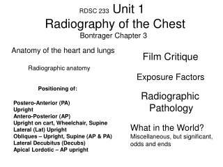
RDSC 233 Unit 3 Radiography of the Upper Gastrointestinal Tract Bontrager chapter 14 Anatomy of the stomach & duodenum Film Critique Radiographic anatomy Exposure Factors Contrast media for the alimentary tract and examination procedures Radiographic Pathology Positioning of: Stomach AP, PA, RAO, LPO, Rt. lateral Motility series What in the World? Miscellaneous, but significant, odds and ends
Atlas of Human Anatomy Second edition (258) Need to know Body habitus relative to position of the stomach and abdominal viscera Centering of the stomach is the pyloric canal/bulb. Note the change in position. Hypersthenic (5%) Sthenic (50%) Hyposthenic (35%) Asthenic (10%)
Atlas of Human Anatomy Second edition (258) Need to know Gastroesophageal junction Cardiac part of Stomach Fundus Lesser and greater curvature Body of stomach Angular notch (incisure angularis) Pyloric antrum & canal Greater omentum Abdominal part of the esophagus
Atlas of Human Anatomy Second edition (259) Need to know Cardiac oriface Gastric (rugal) folds (rugae) Pylorus (sphincter) Superior (first) part of duodenun (ampulla, cap, or bulb) Zigzag (Z) line
Atlas of Human Anatomy Second edition (262) Need to know First (bulb), Second (descendng), third (horizontal), fourth (ascending), parts of the duodenum Duodenojejunal flexure Duodenal papilla (vater) Jejunum Head of pancreas
Atlas of Human Anatomy Second edition (263) Need to know Jejunum Ileum Mesentary Visceral peritoneum (Serosa) Circular folds Mucosa
Radiographic Anatomy Be prepared to identify these anatomical structures in lab.
RAO Position Radiographic Anatomy of the upper gastrointestinal tract Barium in esophagus Rugae (rugal folds) Gas and barium in fundus of S. Left hemidiaphragm Greater curvature of s. Lesser curvature of s. body of s. Incisure angularis Duodenal bulb (or cap) etc. Pyloric canal & Antrum Pyloric orifice (surrounded by sphincter) 2nd or descending part of d. 3rdor horizontal part of d. Jejunum 4th or ascending part of d.
Radiographic Anatomy of the upper gastrointestinal tract Distal esophagus Gastroesophageal junction (at cardiac orifice) Cardiac zone (extends into fundus) Area of detail Left hemidiaphragm Peristalic contraction from distal to abdominal part of esophagus
Anatomy Review Know for lab
Contrast media for the alimentary tract and examination procedures Patient preparation (Prep) Two kinds of Upper GIs Fluoroscopy Spot films Vs. Overheads Contrast media Barium Water soluble iodine preparations The Technologist’s role
Two Kinds of Upper GIs Patient Prep for the Upper GI In the early years of radiology only barium contrast was used. As the film on the left demonstrates, a concentration of barium entirely attenuates the beam. Filling defects appear as lucent shadows in the midst of the barium, or as irregular shapes on the edge of an organ. Calcium or magnesium citrate crystals produce C02 gas, and a translucent, double contrast effect. For the stomach to be visualized free of solids or liquids that could dilute the contrast, or obscure pathology, the patient should be NPO, (Non Per Os), for 8 hrs prior the exam. This restriction includes chewing gum and cigarettes.
Fluoroscopy Spot Films Vs. Overheads Examinations of the alimentary tract begins under fluoroscopy to: * aid in diagnosis (peristalic activity and filling see next screen) Since spot films allow precise positioning, why are overheads even taken? Short answer: Geometric factors. Using the flouro tube there is variable OID, less SID, and therefore more penumbra Digital systems have eliminated overhead filming some places * Identify contraindications to continuing (i.e. fistulas, aspiration of contrast) * Spot filming under fluoroscopic guidance
The effect of positions and barium filling on the visualization of anatomy Identify the common anatomy on these views
Contrast Media for the Alimentary Tract What you need to know Radiopaque (vs. radiolucent) contrast media is typically barium, a soft, metallic, alkaline earth: atomic # 56. Processed into barium sulfate (BaSO4). Salts of barium are chemically pure or they’re poisonous. Insoluble in water. Collodial suspension (shake vigorously). In the GI tract barium is inert. Reactions are extremely rare.
Contrast Media for the Alimentary Tract What you need to know Barium suspensions are tasteless, but rather unpalatable. Flavorings are added to commercial preparations, or may be added when used. Added water changes viscosity and opacity. Thin barium moves though the tract more quickly. Thick barium adheres to the mucosa. Mixtures are defined by department protocols. Water is absorbed by the colon. If a patient is dehydrated, water is absorbed and barium becomes impacted. In addition to drinking water, laxatives, such as castor oil may be given after the exam.
Contrast Media for the Alimentary Tract What you need to know Water soluble contrast is also used to dilute barium preparations s loosing opacity. Barium is contraindicated when there is a chance of leakage (post-surgical, or perforations), and obstruction Water soluble, iodine preparations are absorbed by the body, and used in such cases. Gastroview, Gastrografin, and Oral Hypaque are brand names. All are bitter tasting. Iodine sensitivity is a possibility.
The Technologist’s Role Prepare materials for exam Set up filming equipment Greet the patient, explain exam, take Hx, answer questions Get and introduce radiologist, assist during fluoroscopy. Take and process overhead films Clean up room, enter exam in computer (Meditech at MWMC) Dismiss the patient when radiologist okays
Radiographic Positioning of the Upper Gastrointestinal Tract
Positioning of: AP stomach PA stomach RAO stomach LPO stomach Right lateral stomach and the Motility series including Film Critique
Film Critique Reminder In addition to criteria specific to each projection, all films are evaluated for: * Patient ID * Rt/Lt, special marker * Contrast & density * Motion * Artifacts including
Preparation Standard UGI Positioning 1. Evaluate the order 2. Greet the patient 3. Take History What is pertinent Hx? Abdominal, epigastric, or chest pain (RO/MI),acid reflux, dyspepsia (indigestion), ercuctation, anemia from GI bleed, abdominal mass, N/V, hematemesis. 4. Remove jewelry, check attire, snaps, pins, NG tubes, etc. 5. Explain the exam in layman’s terms 6. Questions? 7. Set technique before positioning
Setup Routine UGI Positioning 1. 40” SID (relatively standard) 2. Reciprocating bucky 12:1, 16:1 grid 3. 100 + kVp for single, and 80 – 90 kVp for double contrast (Why?) 4. 14” x 17” lengthwise for surveys, or 14” x 14” or 10” x 12” for cone down views 5. ID marker generally at bottom 6. Lt or Rt marker free of anatomy.
Routine AP Positioning Steps 1. 14” x 17” 2. Midsagittal plane straight, no rotation 3. CR between xiphoid tip and lower margin of ribs (inferior costal margin) * 1” higher for hypersthenic, * 2” lower for asthenic. 4. Favor left side if needed. 5. Suspend breathing on expiration.
Routine AP Positioning On a 14” x 17” film, the entire stomach, duodenum, and the proximal jejunum (whatever may fill) should be included. Criteria In the supine position the fundus is inferior to the pylorus. Barium fills the fundus, and if double contrast, air fills the body and duodenum. Lateral radiograph, showing position of fundus relative to the pylorus.
Routine PA Positioning Steps 1. In PA position landmarks are less accessible. In addition to 1-2” above lower margin of lateral ribs (Bontrager), 6” above the iliac crestis often used to localize the level of the pylorus and duodenal bulb (center) Higher for hypersthenic, lower for asthenic. 2. Favor left side if needed. 3. Double check Lt. marker (it’s a tricky one)
Routine PA Positioning On a 14” x 17” film, the entire stomach, duodenum, and the proximal jejunum (whatever may fill) should be included. Criteria In the prone position the fundus is superior to the pylorus. Barium fills the Pylorus, duodenum, and if double contrast, air fills the fundus. Lateral radiograph, showing position of fundus relative to the pylorus.
The “Classic” Anterior Oblique Position * Entire spine straight (head elevated) * Coronal plane through shoulders same as coronal plane through hips * Placement of arms and legs like this
The “Classic” Posterior Oblique Position * Entire spine straight (head elevated) * Coronal plane through shoulders same as coronal plane through hips * Placement of arms and legs like this
Routine RAO Positioning The “GI Position” Steps 1. 10” x 12” lengthwise crosswise for transverse stomach or 11” x 14” (dependant on department protocol) 2. 400-700 RAO (steeper for hypersthenic habitus) 3. CR between spine and Lt lateral border, at level of L2. The iliac crest is at the level of the L4-5 interspace. The inferior costal margin of the ribs is at L2-3. 2” higher for hypersthenic, closer to spine, & transverse film 2” lower for asthenic (Observe fluoro)
Routine RAO Positioning The “GI Position” Criteria Of the stomach projections the RAO is the film of choice.In addition to the profile of stomach anatomy, the duodenal bulb and the sweep of the duodenum must be included. Like the PA, air rises to the fundus, and barium settles in the pylorus. Note the marker placement
Steps Routine LPO Positioning 1. 10” x 12” lengthwise crosswise for transverse stomach or 11” x 14” (dependant on department protocol) 2. 300-600 LPO (steeper for hypersthenic habitus) 3. CR same as AP 4. Respiration: same as all Note that the LPO is the corresponding view to the RAO, and yet the degrees of obliquity are 10 degrees less.
Routine LPO Positioning Criteria Like the RAO, the LPO demonstrates the stomach in profile. The duodenal bulb and the sweep of the duodenum must be included. Like the AP, air rises to the pylorus and duodenum, and barium settles in the fundus.
Routine Rt. Lateral Positioning Steps 1. 10” x 12” lengthwise or 11” x 14” (dependant on department protocol) 2. True right lateral position, mid-coronal plane to long axis of table 3. CR to level of inferior costal margain, between anterior bodies of L spine, and muscular wall of abdomen. 4. Respiration: same as all 5. 2” both, for hyper and asthenic habitus
Routine Rt. Lateral Positioning Criteria The Rt. lateral is unique in demonstration of the retro- gastic space. Like the PA and RAO, air rises to the fundus, and barium settles in the pylorus and duodenum. For hypersthenic patients, the Rt. lateral may demonstrate the profile view, pylorus, bulb, and sweep of the duodenum much like the RAO does for other body types.
Review of UGI Film Critique If all the GI views were in a routine, the most efficient order would be: 1. PA 2. RAO 3. Rt. Lateral 4. AP 5. LPO However, for double contrast exams the order may purposefully roll the patient back and forth to keep the mucosal surface coated with barium. On all films Patient ID Rt or Lt marker Contrast & density Motion Artifacts Know where the barium and air will be on the: 1. RAO 2. PA 3. LPO 4. AP 5. Rt Lateral
Motility Series: Chap. 15 (492-493) in Bontrager The small intestine is studied less frequently than other other parts of the GI tract, but when indicated, is usually done in conjunction with the UGI. The examination is known as a motility series or small bowel series (SBS) or small bowel follow through (SBFT) Procedure: Following the UGI, 12 to 16 oz. of thin barium is given the patient. An initial “30 min” KUB is taken, the time is noted, and a schedule of 15 to 30 minute intervals are noted. AP KUBs are taken on this schedule until the barium reached the terminal ilium. The “30 min.” film should be centered higher than a normal KUB. Note the full stomach. Jejunum
Motility Series: Chap. 15 in Bontrager On occasion a PA KUB may be requested by the radiologist. In this position pressure on the loops of bowel may better demonstrate pathology Ileum When the barium reaches the colon, KUB filming is complete. A small bowel series is usually completed with fluoroscopy, and “spotting” the terminal ileum. A compression paddle is used to separate loops of bowel. A metal ring helps center it.
Motility Series: Chap. 15 in Bontrager A SBFT may only take an hour or two. When a patient is very ill, unable to keep down much barium, and laying on a gurney between films, it may take many hours. If the patient must remain recumbent, a Rt lateral or RAO position promotes motility. If the patient is not ill, looking at pictures of food helps speed things along
Enteroclysis: Chapter 15 in Bontrager A method that provides greater visibility of the small intestine is call enteroclysis The patient is intubated with an enteroclysis catheter (Bilbao or Sellink tube), through the nose, directly into the duodenum. High density barium, followed by air or methylcellulose is injected
Exposure Factors From the “Rules of Thumb” Based on: 3 phase, 100 RS film, medium speed screen, 72” SID Compute a technique for a double contrast, and single contrast UGI for a 26 cm abdomen in an AP position, using 400 RS system 1. (2 x 26) + 35 = 87 kVP @ 50 mAs 2. 50 mAs / 4 (RS) = 87 kVp @12.5 mAs (Double contrast) 3. 87 + 13 = 100 kVp @ 6 mAs (Single Contrast) Now put the patient in an RAO (single contrast) Up AP technique by 40 – 60 % = 108 kVp at 6 mAs Now put the patient in a right lateral (double contrast) Up AP kVp by 10, double mAs = 97 kVp @ 25 mAs
Significant Pathologiesof the upper gastrointestinal tractand theirRadiographic Appearances Thoracic stomach Diverticula Ulcerations
Hiatal Hernia & Thoracic Stomach Herniation of the stomach through the esophageal hiatus of the diaphragm. The degree of herniation varys. Sliding hiatal hernia: Small portion of the stomach protrudes though the hiatus due to a weakening of the esophageal sphincter muscle. Herniation changes positions, and may be demonstrated by the valsalva maneuver. Reflux, common to hiatal hernias, may be demonstrated in the trendelenburg position. Schatzke’s ring is a radiographic sign of a sliding hernia. Thoracic stomach: Shown on these images. The majority of the stomach herniates into the thoracic cavity. Thoracic stomach on CXR
Diverticula Diverticula are outpouching caused by weakness in the wall of a hollow organ. They are benign and generally asymptomatic, though the weakened area may perforate, and materials in blind pouches can cause inflammation, ulceration, or infection. This 4 CM diverticula of the first part of the duodenum contains fibrous materials, creating a mass known as a bezoar. Accumulations include hair, cellulose, and seeds Meckel’s diverticulum is caused by a remnant of the yolk sac (umbilical vesicle) found 2’- 4’ from the terminal ileum
Ulcerations Ulcers occur throughout the GI tract, but are most common in the pylorus and duodenum due to the concentration of gastric secretions. These are known as peptic, gastric, or duodenal. A bacteria, H pylori, is a cause of ulcers recently discovered. Radiographically, ulcer craters are seen when barium fills them, and remains after position or peristalsis has moved the rest of the barium on.
What in the World? Miscellaneous, but significant, odds and ends
What in the World? Pathology or Round, radiolucent densities in the GI tract of a boy were very puzzling. They moved, which was good, but what could they be? Other than motion, why might this spot film be so blurry?
What in the World? Is the barium filling in the right hypochondriac/epigastrium region? It starts here
What in the World? How’s it hangin’? Spot films, from the spot film device, may be difficult to identify and hang properly. If they are always in the device the same way the ID marker is used for orientation. If that is unknown the recognition of anatomy is the key. Ribs Vertebral bodies and disk spaces Diaphragm & lung Esophagus Pylorus Cap of d. 2nd part of d. Vertebral bodies and disk spaces Things to look for
What in the World? How’s it hangin’? D B C A Spot films, from the spot film device, may be difficult to identify and hang properly. If they are always in the device the same way the ID marker is used for orientation. If that is unknown the recognition of anatomy is the key. On this and the next two screens, see if you can ID the spot films hung correctly Answers follow