Download
1 / 12
120 likes | 276 Views
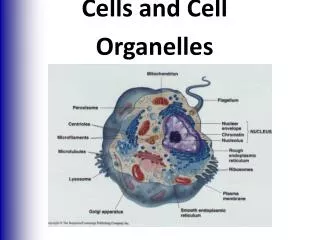
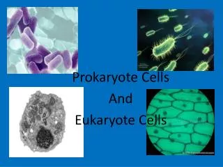
Organelles in Prokaryote cells. P rotists. Most protists are single-celled, some form colonies of cells and some, are multicellular (seaweeds). Are the most diverse of the five kingdoms. They include: fungus-like moulds (slime moulds, downy mildew ),

E N D
Protists • Most protists are single-celled, some form colonies of cells and some, are multicellular (seaweeds). • Are the most diverse of the five kingdoms. They include: • fungus-like moulds (slime moulds, downy mildew), • plant-like organisms (red, green and brown algae, dinoflagellates and diatoms) • animal-like protozoans (flagellates, ciliates, amoebas and sporozoites). • However, protists lack the specialised features usually found in fungal, animal and plant cells (Table 2.1).
Fungi • Some fungi (such as yeasts) are unicellular, but most are made of threadlike filaments called hyphae • Mushrooms and puffballs, the fruiting bodies of some fungi, are made from tightly packed hyphae. • Fungal cells have a cell wall composed of chitin, a substance that also occurs in the exoskeletons of arthropods and is resistant to chemical breakdown. • Fungi are heterotrophs—they must obtain organic molecules by digesting other organisms or their products. Fungi release digestive enzymes, absorb partly digested organic food and store carbohydrate as glycogen, as do animals. • Fungal cells do not have cilia or flagella, therefore fungi cannot move about.
Animals • Multicellularorganisms that are typically highly mobile • Like fungi, are heterotrophic, so their nutrition involves the digestion and absorption of organic food. • DO NOT have a cell wall. Plants • Plants are multicellular, stationary • Autotrophic organisms; they manufacture their own organic materials from inorganic materials using light energy. • Plant cells typically have, in addition to the features seen in animal cells, a cell wall made of cellulose
Viruses • Subcellulargenetic parasites. • Not classified as living organisms and they are not cells. • Capable of organising their replication and can direct the construction of a surrounding protein coat. • They are completely parasitic and multiply within host cells using the host cells’ materials and processes, often killing the cells in the process. • Viruses reproduce only inside the cells of a host organism, and infect bacteria, protists, fungi, plants and animals. • Egs common cold influenza, AIDS, mumps, measles, rabies and smallpox in humans, and myxomatosisin rabbits. Polyviruses infect many plants, including potatoes. The viruses that infect bacteria are called bacteriophages (meaning ‘bacteria eaters’) (Figure 2.15). • A single virus particle (virion) is composed of genetic material (DNA or RNA) enclosed in a protein coat known as a capsid. Viruses do not have cytoplasm, membranes or any organelles. Some viruses have additional outer layers of protein, and may become wrapped in host plasma membrane as they bud out of their host cell.
Investigating Cells Light Microscope • Each of these uses visible light (electromagnetic radiation in the visible range) to examine cells and tissues. • The preparation of tissue for microscopy involves several steps that are the same, in principle, for both light and electron microscopy. • First, the tissue is ‘fixed’; that is, the various parts of a cell are chemically ‘immobilised’ so that the structure of the cell remains as lifelike as possible. • Next the tissue needs to be sliced into thin slices to allow light to pass through • Different stains are used to highlight different parts of the cellular organelles
Types of light microscopy • Autoradiography • Fluorescence microscopy • Confocalmicroscopy
Electron Microscopy • In electron microscopy, an object is viewed using an electron beam instead of light. This allows a much higher resolution (ability to see fine detail), • Athousand times greater than light microscopy. • The general steps of electron microscopy include fixation, embedding, sectioning and staining. • For transmission electron microscopy (where the electron beam travels through the section), fixation must be rapid and precise because very fine details of cellular structure are examined. • Electrons absorbed or deflected by the electron dense ‘stain’ will not pass through, leaving a shadow on the screen lit by those electrons that are transmitted. Electron microscopy produces only black and white images (although these may later be coloured). • Scanning electron microscopy also uses electrons instead of light, but instead of transmitting them through the specimen, the electrons are bounced off a three-dimensional surface of fixed tissue that has been coated with metal (gold). This gives a high resolution shadow picture of the surface features.
Synchrotron • A synchrotron is basically a big circular tube inside which electrons are travelling at close to the speed of light. Synchrotron light is the very bright electromagnetic radiation emitted as these charged particles are forced to change direction under the action of a magnetic field. The electromagnetic radiation is emitted in a narrow cone in the forward direction, at a tangent to the particle’s orbit. • Visible light occupies just a small part of the electromagnetic spectrum. • Synchrotron light is unique in its intensity and brilliance, and it can be generated across the range of the electromagnetic spectrum • Synchrotron light allows scientists to explore matter in ways that were unimaginable a few years ago. • Synchrotron light allows matter to be ‘seen’ at the atomic scale, including the nanosecond-by-nanosecond behaviourof protein molecules such as antibodies. It enables scientists to collect, in hours, data on the structure of proteins that would once have taken weeks or months. While structural biology is their most important application, synchrotrons are useful in many other areas, such as nanotechnology and materials science. • Until now Australian scientists have had to use used synchrotrons at overseas facilities. The new Melbourne synchrotron allows complex protein structures to be determined quickly and is central to drug design and development. It allows further development of medical imaging technologies, such as phase contrast microscopy. Synchrotron analysis of biological samples can also help to identify diseases. For example, Australian research has suggested that a single hair from a woman could reveal whether she has breast cancer.