Download
1 / 36
370 likes | 536 Views
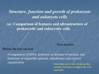
Visualizing Prokaryote Cells. Chapter 3 - Black. Light. Key Words - . Visible light Ultraviolet light Reflection Transmission Refraction Absorption. Light Microscopy. Resolution. Resolution is the ability to see two images as separate and discrete.

E N D
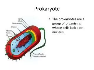
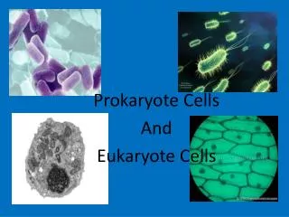
Visualizing Prokaryote Cells Chapter 3 - Black
Key Words - • Visible light • Ultraviolet light • Reflection • Transmission • Refraction • Absorption
Resolution • Resolution is the ability to see two images as separate and discrete. • The wavelengths of visible light from 420 to 620 prevent resolution of two points closer than 220 nm • By using the light emitted from an electron it is possible to resolve two points that are .2nm apart
Specialized Microscopy • Dark field • Phase Contrast • Differential interference • Fluorescence • Confocal • Digital
Digital photomicrosopy • Camera can be used to photograph images • Specimen can be viewed on TV screen as well as on computer screen
Phase Contrast Microscopy • Accentuate small differences in the refractive index of the specimen • More detail is apparent in living cells • Assist in the visualization of cell structure in transparent cells • micro.magnet.fsu.edu/.../dicphasecomparison.html
Differential interference microscopy • Produces higher resolution • Depends on a gradient • It can produce almost a three dimensional image
Comparison of Phase Contrast and Differential Interference Contrast
Fluorescent Microscopy • Organisms such as Mycobacterium tubercuolosis and Treponema pallidum are treated with a fluorochrome dye • Ultra violet light is used to excite the fluorochromemolecules and produce a glowing image • Used in clinical work • Also used with antigens and antibodies to identify the presence of molecules on the surface of a cell
Fluorescent antibody staining • Used in immunology • Fluorescent antibodies are used to detect antigens on the surface of cells • In the picture to the right are Bacillus anthracis cells tagged with a fluorescent antibody
Confocal Microscopy • Utilizes beams of ultraviolet light to excite fluorescent dye molecules. • The exciting light is focused on the specimen with a thin optical fiber • Resulting fluorescence is focused through a narrow aperture • The light is detected and analyzed by a computer • Very sharp focus • For thick specimens an image is constructed in layers
Confocal images • http://www.microscopyu.com/galleries/confocal/applecedarrustaeciaandpycnia.html • http://www.microscopyu.com/galleries/confocal/chlamydomonas.html
Electron Microscopy • Transmission electron microscopy • Scanning electron microscopy
Transmission Electron Microscopy • Electrons are used as the source of light • Produced by a high voltage current running through a tungsten filament • www.steve.gb.com/science/electron_microscopy.html
Transmission Electron Microscope • The lenses are electromagnetic • They act on the negatively charged electrons to focus them in a concentrated path through the specimen • The image is magnified by additional lenses and visualized on a screen
TEM images • Images produced display high resolution • Staining with heavy metals that interact with the electrons • Gradations of black, gray, and white contrast areas of greater density that absorb the stain
Scanning Electron Microscopy • Electrons are reflected and collected off of the surface of a cell
SEM • Images show surface contours • Three dimensional image
Freeze Fracture • Cells are quickly frozen in liquid nitrogen (196C), which immobilizes cell components instantly.2. Block of frozen cells is fractured. This fracture is irregular and occures along lines of weakness like the plasma membrane or surfaces of organelles.3. Surface ice is removed by a vacuum (freeze etching)4. A thin layer of carbon is evaporated vertically onto the surface to produce a carbon replica.5. Surface is shadowed with a platinum vapor.6. Organic material is digested away by acid, leaving a replica.7. Carbon-metal replica is put on a grid and examined by a transmission electron microscope.
References • http://micro.magnet.fsu.edu/primer • http://www.mos.org/sln/SEM/