Download
1 / 122
1.22k likes | 1.41k Views
Nephrotic syndrome (NS) 肾病综合征. Department of Pediatrics Soochow University Affiliated Children’s Hospital. Concept. Heavy proteinuria Hypoproteinaemia Oedema Hyperlipidaemia. Physiology. Heavy proteinuria---reasons. Injured glomerular filtration barrier. 足 突 细 胞. 足 突 细 胞 的 超 微

E N D
Nephrotic syndrome (NS)肾病综合征 Department of Pediatrics Soochow University Affiliated Children’s Hospital
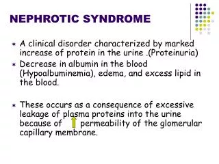
Concept • Heavy proteinuria • Hypoproteinaemia • Oedema • Hyperlipidaemia
Heavy proteinuria---reasons Injured glomerular filtration barrier
足 突 细 胞
足 突 细 胞 的 超 微 机 构
Heavy proteinuria--results ? Prolonged glomerular leakage of protein leads to hypoalbuminaemia ,that is associated with loss of fluid into the extracellular space, Reduction in circulating volume stimulates renal retention of salt and water, manifested as oedema.
Hypoalbuminaemia-reasons? • Urinary losses of albumin are the major factor in the development of hypoalbuminaemia • possible role of catabolism of reabsorbed albumin in the tubule
Hyperlipidemia: • Due to Increased Hepatic synthesis • Decreased Large molecular Lipidoprotein depletion • high LDL, TG, cholesterol and low HDL level cause cardiovascular diseases • At same time it also lead to glomerular sclerosis and tubulointerstitial fibrosis
Oedema—reasons? • Reduced oncotic pressure • Low blood volume • Increased Na and water absorption • Increased RAS activity • Decreased GFR
Others physiologic changes • Reduced immunity : low IgG • High coagulative status : thrombi • Lost of Microelements : Ca, Zn, Fe, Copper • Vitamin D deficiency • Hormone: T3, T4
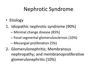
Pathology • Idiopathic NS 76.4% ( Nil disease, Lipoid Nephrosis, Minimal change disease, MCD ) • Focal Segmental Glomerular sclerosis (FSGS) 6.9% • Mesangial Capillary proliferative GN ( Membranous Proliferative GN, MPGN)7.5% • Mesangial GN (MsPGN)2.3% • Membranous GN (MN) 1.5%
Diagnosis (1) • Clinical :congenital , primary or secondary simple or nephritis NS • Pathologic: minimal changes and non minimal changes • Therapeutic : steroid sensitive , steroid resistance , steroid dependence and frequency relapse NS
Diagnosis(2) • Congenital NS • Primary NS 90% • Secondary NS 10%: HSP; SLE, IgAN, vasculitis, Wilson’s disease, tumor disease,
Diagnosis (3) Simple NS : Nephritis NS : 3 highs and 1 low 1 high and 1 low with any one item below Haematuria : no >10/HP within 2 weeks Hypertension: no Yes BUN & SCr: normal Persistence Increased : C3 : normal Low
Treatment general therapy 1. Have a rest : don’t need enforced bed rest other than severe oedema and hypertension 2. Infections: prevention, prophylaxis therapy; vigor treatment; 3. vaccine? 4.Diuretics: combined potassium keeper
Diet: 1.low salt and water diet need only in severe oedema and hypertension, medium protein input (1.5-2g/Kg) is suggested 2. high protein feeding merely led to increased spilling of albumin into the urine without an increase in plasma albumin concentrations (Kaysen et al. 1989). 3. low protein diets may lead to negative protein balance in the long term
Treatment (2) Specific treatment Steroid : prednisolone: first selection dexmethasone methylprednisone Principle: dose must be up to 2 mg/Kg.d maximum, 60mg/d; taper slowly; medium-long term maintenance
Steroid---term • Short term therapy : 8 weeks: • Medium term therapy: 6 months • Long term therapy: >9 months
Therapeutic judgment • Steroid Sensitive NS (SSNS) • Steroid Partial Sensitive NS • Steroid Resistant NS (SRNS) • Steroid Dependent NS (SDNS) • Frequent Relapse NS
Side effects Short terms: predisposing infections electrolyte unbalance Lon terms : cohn’s syndrome bone malnutrition diabetic cardiovascular: eyes problem:cataract psychological disease:
Cytotoxic drugs Indications : steroid resistant NS steroid dependent NS frequently relapse NS severe pathologic change NS: FSGS
Cyclophosphamide • Mechanism: DNA mitosis level • Side effects: gonad damage • Administration: Oral , Intravenous pulse
others • Chlorambucil : mitosis level • Azathiopurine : synthesis level • Cyclosporine or FK 506 : activation T cells level • Mycophenolic acid : synthesis level • VCR: synthesis level
Anti-coagulations Anti—coagulation factor :heparin Anti—platelet accumulation : dipyridamole Fibrin split drugs : urokinase
Immune regulation drugs • Levamisole • Intravenous Immunoglobulin • Chinese herbs
Prognostic outcome • Simple nephrotic syndrome: good: minimal change nephrotic syndrome infections and malnutrition • Nephritis nephrotic syndrome: poor: nonminimal change nephrotic syndrome, renal malfunction , hypertension, low complement
Homework • The management of urinary tract infections • The investigations of a child with proteinuria
You should have an understanding of : • The investigation of a child with haematuria • The basic symptoms and signs suggestive of renal failure, and have a working overview of its management.
Concept • Urine 10 ml 1500rpm spin for 10 minutes . 0.2ml precipitation under microscope • Pathological: More than 3 red cells per high field under microscope • As a result of disease : kidneys and lower urinary tract, as well as coagulopathies, drugs and exercise
Glomerular causes of haematuria • Glomerularonephritis • Recurrent haematuria : IgA nephropathy • HUS: • HSP: • SLE:
Non-glomerular • Polycystic diseases • Congenital anomalies • Infection • Trauma • Tumors • Calculi : hypercalciuria • Nut cracker syndrome
Non renal causes • Haematological : Coagulopathies; thrombocytopenia; renal vein thrombosis; sickle cell diseases • Non-haematological: exercise; Drugs
Glomerulonephritis(GN): Poststreptococcal GN • Is the most common form of Glomerulonephritis and caused by certain strains of streptococci (group A -hemolytic streptococcal infection • It affect young children greater than 3 yrs of age • The incidence is declining due to reduced infection rate and prophylactic antibiotics • Sex: More boys than girls ,about ratio of 2: 1
Pathology(2) • The glomerular tuft shows an increased number of cells and appears to fill up the Bowman’s space: mesangial , endothelial, and epithelial cells proliferate ( crescent GN) • There is infiltration of cells from the circulation: Polymorphonuclear leukocyte; Monocytes and T lymphocytes • Immune deposits in the glomerular basement membrane and mesangium can be seen by immunofluorescence microscopy. • Electron-dense subepithelial deposits (humps) are typical of acute poststreptococcal glomerulonephritis