Download
1 / 23
261 likes | 762 Views
Nephrotic nephritic syndrome. By Dr Rasol M Hasan. Here’s one of those things in pathology that will lead you to pull all your hair out: what is the difference between nephrotic and nephritic syndrome ? Ugh. They both involve the kidney, they both

E N D
Nephrotic nephritic syndrome By DrRasol M Hasan
Here’s one of those things in pathology that • will lead you to pull all your hair out: what is • the difference between nephrotic and • nephritic syndrome? • Ugh. They both involve the kidney, they both • are syndromes so they’re probably both • constellations of findings, and the names are • maddeningly similar except for one stinking • vowel. How can a person be expected to • memorize these things?
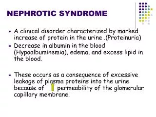
Nephrotic Syndrome • The patient will present with a triad of symptoms: • Proteinuria , i.e. >3g/24hr-3.5g/24 hr • Hypoalbuminaemia, i.e. <30g/L • Oedema
Capillary Space Filtration Membrane – Electron Micro. MD consult GBM Endothelium Urinary Space Podocyte 4
To avoid overlooking this diagnosis, carry out a urine dipstick test on all patients with oedema, looking for proteinuria. • >80% of cases are due to glomerulonephritis.
In this syndrome, there is damage to podocytes. It was once thought that this allowed albumin to leak out into the tubule, thus causing proteinuria and hypoalbuminemia, and leading to reduced plasma oncontic pressure and peripheral oedema. The damage to the podocytes was thought not to be significant enough to allow RBC’s through the gaps thereby rendering haematuria unlikely.
Recently, this theory has come under scrutiny as it has been discovered that the aforementioned situation does not cause a change in oncotic pressure, confirming the presence of an alternative pathology causative of oedema.
Clinical signs • Pitting oedema, particularly in the limbs and around the eyes; may also cause genital oedema and ascites. • Possible hypertension
Causes • Primary causes – these are diagnoses of exclusion that are only made if secondary causes cannot be found • Minimal change disease (MCD) • Focal segmental glomerulosclerosis • Membranous nephropathy
Secondary causes – note that these fall into the same three categories as above: • Minimal change disease – Hep B, SLE, diabetes M, sarcoidosis, syphilis, malignancy • Focal segmental glomerulosclerosis –HIV, obesity, diabetes M, hypertensive nephrosclerosis • Minimal change disease –drugs, malignancy, particularly Hodgkin’s lymphoma
Differential diagnoses include cardiac failure, i.e. increased JVP, pulmonary oedema and mild proteinuria, and liver disease, i.e.reduced serum albumin. • The condition causesan increased susceptibility to infection – partly due to loss of immunoglobulin in the urine. Patients tend to be prone to streptococcus infection, as well as bacterial peritonitis and cellulitis. • Nepthrotic syndrome also increases the risk of thromboembolism and hyperlipidaemia.
The former is due to an increase in the synthesis of clotting factors and to platelet abnormalities, and the latter is a result of increased synthesis of these by the liver to counteract reduced oncotic pressure.
Investigations • These are the same as those carried out in GN. • Also, check for cholesterol as part of confirming the presence of hyperlipidemia. • Renal biopsy – order this for all adults. In children, because the main cause is minimal change GN, steroids are the first-line treatment. Therefore, in children, biopsy is necessary only if pharmaceutical intervention fails to improve the situation.
The hypercoagulant state seen in the nephrotic syndrome can be a risk factor for renal vein thrombosis. This can present as loin pain, haematuria, palpable kidney and sudden deterioration in kidney function. This should be investigated with Doppler USS, MRI or even renal angiography. • Once diagnosed, give warfarin for 3 to 6 months.
Management • Generally, this involves treatment of the underlying condition which is usually GN. Therefore, fluid management and salt intake restriction are priorities. The patient is usually given furosemide along with an ACE inhibitor and/or an angiotensin II receptor antagonist. Prophylactic heparin is given if the patient is immobile. Hyperlipidaemia can be treated with a statin.
Nephritic Syndrome • Again, this is a term describing a set of symptoms and not a pathological condition in itself. Acute and chronic forms of the syndrome exist. The main difference between this and nephrotic syndrome is that in nephritic syndrome haematuria is present. There is also proteinuria, hypertension, uraemia, and possibly oliguria. The two standout features are hypertension and RBC casts. The urine will often appear ‘smoky’ in colour due to the presence of RBC casts. Very rarely, it may appear red
Causes • Post-streptococcal • Primary: • Membranous glomerulonephritis • Rapidly progressive glomerulonephritis • IgA nephropathy (Berger’s disease) • Secondary • HSP • Vasculitis
CLINICAL FEATURES • Abrupt onset of : • glomerularhaematuria (RBC casts or dysmorphic RBC). • non-nephrotic range proteinuria ( < 2 g in 24 hrs) . • oedema ( periorbital, sacral ). • hypertension. • transient renal impairment (oliguria, uraemia).
Urinary casts – these are cylindrical structures produced by the kidney and present in the urine in certain renal diseases. They form in the DCT and collecting duct, dislodging and passing in the urine where they are detected by microscopy. RBC casts are usually associated with nephritic syndrome. The presence of RBCs within a cast is always pathologic and strongly indicative of glomerular damage.
The proteinuria present is often smaller than in nephrotic syndrome, thus a coexistent condition of nephrotic syndrome is not usually present.
Encepelopathy may be present, particularly in children, due to electrolyte imbalances and hypertension. This type of presentation is indicative of glomerular damage, but requires renal biopsy to determine the exact problem. In this respect it is similar to nephrotic syndrome. • Overlapping of the two syndromes is possible as nephrotic syndrome may precede nephritic syndrome, although not vice-versa.
Mechanisms of the syndrome vary according to cause; both primary and secondary causes exist. Post-infectious GN is the classic illustration of nephritic syndrome, but the condition may be caused by other glomerulopathies and by systemic diseases such as connective tissue disorders