Download
1 / 9
90 likes | 288 Views
Two Distinct Actin Networks Drive the Protusion of Migrating Cells. A. Ponti, M. Machacek, S. L. Gupton, C. M. Waterman-Storer,*† G. Danuser*†. Proceso de migración celular: Protusión de membrana plasmática en el frente de avance Formación del sitio de adhesión detrás de la protusión

E N D
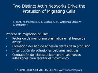
Two Distinct Actin Networks Drive the Protusion of Migrating Cells A. Ponti, M. Machacek, S. L. Gupton, C. M. Waterman-Storer,*† G. Danuser*† Proceso de migración celular: • Protusión de membrana plasmática en el frente de avance • Formación del sitio de adhesión detrás de la protusión • Interrupción de adhesiones celulares antiguas • Contracción del citoesqueleto contra las nuevas adhesiones para facilitar el movimiento 17 SEPTEMBER 2004 VOL 305 SCIENCE www.sciencemag.org
Quantitative Fluorescent Speckle Microscopy (qFSM) • Monómeros de actina marcados fluorescentemente. • Polimeración de un grupo de monómeros formando f-actina (2-8) emiten puntos de fluorescencia, llamados speckles o “brillos” • Podemos estudiar la organización de f-actina en células migrando. 17 SEPTEMBER 2004 VOL 305 SCIENCE www.sciencemag.org
Flujo de f-actina y recambio (turnover) de f-actina 17 SEPTEMBER 2004 VOL 305 SCIENCE www.sciencemag.org
Transición lamela-lamelipodio 17 SEPTEMBER 2004 VOL 305 SCIENCE www.sciencemag.org
Clases de “brillos” 17 SEPTEMBER 2004 VOL 305 SCIENCE www.sciencemag.org
Características moleculares de las redes • Blebistatina: Inhibe actividad adenosin trifofatasa de miosina II no muscular. • Citocalasina D: Inhibe polimerización de terminaciones “barbed”. • Jasplakinólido: Inhibe la despolimerización 17 SEPTEMBER 2004 VOL 305 SCIENCE www.sciencemag.org
Tratamiento con Citocalasina-D 17 SEPTEMBER 2004 VOL 305 SCIENCE www.sciencemag.org
Adhesiones focales al sutrato GFP-vinculina (blanco) 17 SEPTEMBER 2004 VOL 305 SCIENCE www.sciencemag.org
Conclusiones • Dos redes en el borde de las células epiteliales: lamela y lamelipodio. • Estas redes se superponen y están acopladas por enlaces débiles • La transición entre ellas está definida por el comienzo de uniones sustrato-citoesqueleto • La lamela es requerida para el avance productivo de la célula • El lamelipodio probablemente posea funciones exploratorias 17 SEPTEMBER 2004 VOL 305 SCIENCE www.sciencemag.org