Download
1 / 19
190 likes | 433 Views
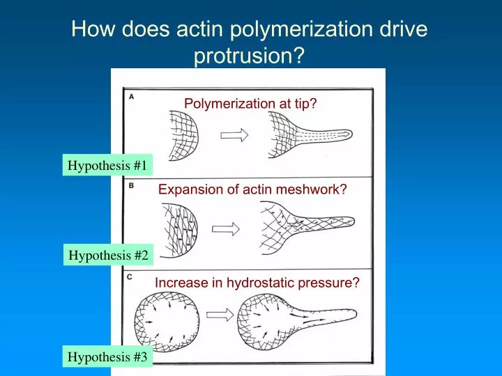
Polymerization at tip?. Expansion of actin meshwork?. Increase in hydrostatic pressure?. How does actin polymerization drive protrusion?. Hypothesis #1. Hypothesis #2. Hypothesis #3. Evidence for #1: The Acrosome reaction. Stages during fertilization of a sea-urchin egg.

E N D
Polymerization at tip? Expansion of actin meshwork? Increase in hydrostatic pressure? How does actin polymerization drive protrusion? Hypothesis #1 Hypothesis #2 Hypothesis #3
Evidence for #1: The Acrosome reaction Stages during fertilization of a sea-urchin egg Elongation of the acrosomal process results from a burst of actin polymerization at the tip. This allows the sperm to penetrate the jelly coat surounding the egg
Evidence for #2: Gel Swelling mechanism of protrusion • 1. Protrusion in Dictyostelium starts as a bleb and actin fills in behind. • 2. During the acrosome reaction: • Increased osmolarity, decreases rate of acrosomal actin filament elongation. • Decreases in osmolarity, increase rate of acrosomal actin polymerization.
Evidence for #3:Myosin I driven protrusion Actin filament sliding mechanism of protrusion Myosin I at leading edge • Myosin I “walks” toward + end while associated with the plasma membrane • Actin filaments slide rearwards, relative to membrane • This may provide space for actin monomers to add to + ends
To understand how actin polymerization drives protrusion we need to know: • 1. Where the nucleation of actin filaments occurs • 2. How high rates of actin polymerization are maintained at the protruding edge • 3. How polymerization generates a protrusive force • To be covered later in this course
APBs involved in regulating actin dynamics Lodish 5th Ed. Chapter 19, p786-791 • 1. Dynamics • Thymosin -4 (G-actin sequesterer) • Profilin (Increases rate of polymerization) • Gelsolin (Increases rate of actin filament turnover) • Capping proteins (Increases rate of polymerization) • Arp2/3 (Nucleation )
Proteins in the cytoplasm sequester or bind to actin monomers - preventing them from polymerizing. Factors which influence the binding of these proteins to actin monomers will affect the rate of actin polymerization. Active sequesterers Inactive sequesterers Thymosin -4 and Profilin are monomer sequestering proteins • Fact: The Cc for actin filament polymerization is 0.1uM, the total actin concentration in a cell is 200uM. 40% of actin in cells is unpolymerized. Why ?
Microinjection of excess TB4 into cells causes loss of stress fibers • Although actin stress fibers are relatively stable turnover of actin monomers is occurring. • Monomers leaving a stress fiber will be rapidly sequestered by TB4. Gradually the stress fiber will disappear. • The equilibrium is shifted toward increasing monomer concentration at the expense of f-actin. After Before
Profilin increases the rate of actin polymerization • Profilin binds to actin opposite the ATP binding cleft* • * allows exchange of ADP for ATP, contrasts with T-4 • Profilin-actin complex to binds readily to the + end of the actin filament (affinity of complex > than single actin monomer • A conformational change in the complex occurs after binding to +end actin filament, causing profilin to fall off
Profilin competes with T-4, for actin monomers • When a small amount of profilin is activated it completes with thymosin for G-actin and rapidly adds it to the +end of F-actin • The activity of profilin is regulated by: • phosphorylation, binding to inositol phospho-lipids • The activity of profilin is increased close to the plasma membrane by binding to: • acidic membrane phospholipids, certain proline rich proteins that localize at the plasma membrane
Functions of the actin cytoskeleton dependent on polymerization • The acrosome reaction. • The rapid formation of an acrosomal process penetrates the thick jelly coat of the sea urchin egg allowing nuclear fusion between sperm and egg. • Before fertilization short actin filaments lie in a pocket at the head of the sperm together with many profilin-actin complexes • Upon contact with the egg, the acrosomal vesicle is exocytosed, uncovering + ends of actin filaments. • At the same time, profilin (of the profilin-actin complex) is activated resulting in the rapid addition of G-actin to the exposed +ends of the pre-existing actin filaments • This results in an explosive elongation of the acrosomal process • The acrosomal process contacts the egg plasma membrane and fuses with it. • The sperm and egg nuclei fuse.
Conclusion: • Newly polymerized actin is found at the leading edge. Only newly polymerized actin is labeled Experiment to demonstrate the location of newly polymerized actin • 1. A fibroblast was microinjected with rhodamine (red) labeled actin monomers • 2. Cell was fixed shortly after microinjection. • 3. The cytoskeleton was stained with fluorescein phalloidin (green). • New actin polymerization occurs within the actin cortex that lies just beneath the plasma membrane • Actin polymerization in this location can form a variety of surface structures • Microvilli, filopodia, lamellipodia • Nucleation of actin filament growth is regulated by external signals • Nucleation is initiated by a comlex of 7 proteins called the ARP2/3 complex All actin is labeled in a lamellipodium
The role of Arp2/3 in protrusion • Arp2/3 is a highly conserved complex of 7 proteins, including 2 actin related proteins (Arp2 and Arp3) • Identified first in the cortical (submembranous) actin of amoebae • Found in highly dynamic actin structures in many cell types • e.g. Listeria (actin tails), edge of lamellipodia, cortical actin patches (yeast)
Arp2/3 nucleates actin filament assembly • Arp2/3 is present at high (~ 10uM) concentrations in motile cells e.g. leukocytes • Arp2 and Arp3 are 45% similar to actin monomers
Arp2/3 provides a “template” for actin filament growth • Arp2/3 nucleates actin filament by binding to the - end of the actin filament • Arp2/3 can bind to the sides of pre-existing actin filaments, resulting in the development of a branching mesh of actin filaments • Nucleation is more efficient when ARP2/3 is bound to the side of an actin filament
Arp2/3 Keratocyte Immunogold labeling of Arp2/3 Actin Overlay Distribution of Arp2/3 in a moving cell Svitkina and Borisy 1999
How is Arp2/3 activated • WASp, Wiskott-Aldrich Syndrome protein, mutated protein leads to bleeding, immunodeficiency - is rich in proline • WASp is activated when it binds PIP2 and active Cdc42 (small GTPase) • VCA domain of WASp is necessary for Arp2/3 activation - binds actin and ARP2/3, --increases affinity of ARP2/3 to side of filament • Other (proline rich) activators of ARP2/3 include VASP (Vasodilator-stimulated phosphoprotein), Scar/WAVE family proteins
A conformational change occurs when Arp2/3 activated • VCA domain of WASp becomes more compact when bound to G-actin • A conformational change occurs so that ARP2 and ARP3 move closer together, to form a template for actin filament growth • In budding yeast and Dictyostelium Myosin I may bind (via SH3 domains) ARP2/3 – possibly transporting it to the protruding edge.