Download
1 / 38
380 likes | 706 Views
Circulatory & Respiratory System. By: Alica Boleware Grace Oh Daniel Naji. The Respiratory System.

E N D
Circulatory & Respiratory System By: Alica Boleware Grace Oh Daniel Naji
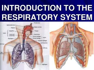
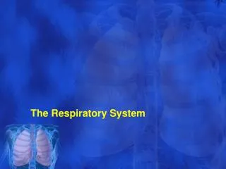
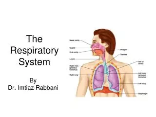
The Respiratory System • The respiratory system is situated in the thorax, and is responsible for gaseous exchange between the circulatory system and the outside world. Air is taken in via the upper airways (the nasal cavity, pharynx and larynx) through the lower airways (trachea, primary bronchi and bronchial tree) and into the small bronchioles and alveoli within the lung tissue.
Respiratory System: The Anatomy of the Nose • The nose consists of certain parts, such as: external meatus- triangular-shaped projection in the center of the face; external nostrils - two chambers divided by the septum; septum - made up primarily of cartilage and bone and covered by mucous membranes. The cartilage also gives shape and support to the outer part of the nose; nasal passages- passages that are lined with mucous membranes and tiny hairs (cilia) that help to filter the air.
Respiratory System:The Anatomy of the Pharynx • Pharynx can be divided into three regions: nasopharynx, oropharynx, and the laryngopharynx.
The Pharynx:The Anatomy of Nasopharynx • Continuous with the nasal cavity via the internal nares. • Lined by respiratory epithelium. • Contains the opening to the auditory tube, also called the Eustachian Tube, which connects the pharynx to the middle ear cavity. • It functions to ensure that the air pressure within the middle ear cavity is equal to atmospheric pressure. • Contains the pharyngeal tonsil.
The Pharynx:The Anatomy of Oropharynx • Oropharynx • Inferior to the uvula and superior to the epiglottis. • Lined by non-keratinized stratified squamous epithelium due the fact it is a common pathway for food and air. • The palatine tonsils are located near the opening of the oral cavity into the pharynx.
The Pharynx:The Anatomy of Laryngopharynx • Laryngopharynx • Inferior to the epiglottis and superior to the split between the larynx and the esophagus. • Similar to the oropharynx, it is lined by non-keratinized stratified squamous epithelium because it is a common passageway for food and air. • Lingual tonsils are located on the posterior surface of the tongue, which also places them near the opening of the oral cavity into the pharynx
The Respiratory System:The Anatomy of the Larynx • The Larynx routes food and air down their correct passages. Larynx has an arrangement of 9 cartilages connected by membranes and ligaments and lined by respiratory epithelium. Certain kinds of cartilages (all hyaline expect epiglottis), such as: thyroid, cricoid, epiglottis (made up of elastic cartilage) and 3 small paired cartilages. Thyroid cartilage is the largest and its midline laryngeal prominence, which is the famous male’s “Adam’s apple.” The 3 pairs of small cartilages form much of the posterior and lateral larynx. The epiglottis extends from the base of the tongue to its hinge on the superior thyroid cartilage.
The Respiratory System:The Anatomy of Trachea • The trachea extends from the larynx to the mediastinum, where it splits into 2 primary bronchi that are lined by respiratory epithelium which is also associated with abundant mucus secretion. • The trachea is reinforced by about 18 C-shaped rings of cartilage. These rings prevent the trachea from collapsing during inspiration. The open portion of the cartilage rings is posterior and there you find the trachealis muscle. The lack of posterior cartilage is significant due to the fact it provides the esophagus with room to expand when a large bolus of food is swallowed. • The trachea has a carina which is the point where the trachea divides into the left and right primary bronchus.
The Respiratory System:The Anatomy of the Bronchi • Bronchi is divided into two main bronchi; the left and the right. It is the air entrance to each lung. • Bronchioles are the smaller airways that sends air to the inside walls of lungs where the alveoli is.
The Respiratory System:The Anatomy of the Lungs • The lungs are divided into lobes; The left lung is composed of the upper lobe, the lower lobe and the lingula (a small remnant next to the apex of the heart), the right lung is composed of the upper, the middle and the lower lobes. • Alveoli is located inside walls of the lungs; it allows the oxygen to be absorbed by the blood cells and oxygenates the blood for transfer throughout the human body. It is also the site of gas exchange in the lungs, must be moist for gases to diffuse.
The Respiratory System:The Anatomy of the Diagraphm • Diaphragm is a layer of muscles that is located at the bottom of the chest. It functions to help pump the CO2 out of the lungs and pull the O2 into the lungs. When the diaphragm contracts, oxygen is pulled into the lungs; when it relaxes, carbon dioxide is pumped out of the lungs
Passage of Oxygen Gas Sinus Pharynx Larynx Trachea nose Bronchi Bronchioles Circulatory Alveoli
The Respiratory System: Key Terms • Nose is the organ of smell that enters air into the animal’s body; it functions to prevent any airborne particles proceeding into the lungs. • Pharynx is known as the throat; it extends from the mouth to the larynx. • Larynx is known as the “voice box”; it functions as an airway to the lungs. • Trachea is the cartilage rings, strong but flexible. It is lined with ciliated cells that push mucus and foreign particles upward.
Mechanics of Breathing Exhalation Diaphragm relaxes (moves up) Diaphragm contracts (moves down) Inhalation Inhalation Exhalation diaphragm
Respiratory Issues:Asthma • Asthma is a chronic disease that affects 300 million people worldwide. • Symptoms include repeated attacks of loss of breath and wheezing. • Asthma can be caused by: • Genetics • Environment (pollution) • Allergens (cat hair, dander, pollen, mold) • Smoke • Asthma can have various triggers: • Cold air • Some medications • Emotional stress
Respiratory Issues:Bronchitis • Bronchitis is an inflammation of the bronchial tubes, which carry air to the lungs. • As a result, excess mucus is produced, inducing coughing fits. • Acute Bronchitis is a short-lived affliction with a quick onset and recovery, with only about 2-3 weeks of symptoms. • Chronic Bronchitis is a long-term affliction that mostly plagues smokers. • Chronic Bronchitis is characterized by a period of at least 3 months of symptoms for a minimum of two years.
Respiratory Issues:Pneumonia • Pneumonia is an infection of the lungs that causes coughs, fever, and breathing difficulties. • It can be caused by viruses, bacteria, or parasites. • Symptoms include: coughing, fever, chest pain, chills, and fatigue. • Chronic diseases such as asthma, cancer, diabetes, and heart problems increase your risk. • Diagnosed by an X-ray or blood test.
What Is Next? • Between the lungs, the heart fits snugly in the middle of the chest leading us to the circulatory system.
The Circulatory System • The circulatory system is made up of the vessels and the muscles that help and control the flow of the blood around the body. This process is called circulation. The main parts of the system are the heart, arteries, capillaries and veins.
The Circulatory System: The Anatomy of the Heart • The center of the circulatory system is the heart, which is the main pumping mechanism (for blood). The shape of the heart is described as a cone, with a pointed bottom and round top. Apparently, the hollowness of the heart is for has the ability to be filled with blood. The heart is made of a muscular wall called a septum - solid enough for the blood to have the inability to flow back and forth between the left and right halves of the heart.
The Heart is in Half? • The heart is broken into four chambers - each top chamber is called an atrium, "the holding chamber" while the bottom chambers are called ventricles, "the pumping chamber". Hence, each side of the heart has its own system - a right heart and a left heart. Each side consist of an atrium and a ventricle while blood can flow from the top chamber to bottom chamber but never between both sides. Because the blood can flow from the atria down to the ventricles, there are openings, called valves, in the walls to separate them. Valves have the ability to open in one direction to let blood pass through, however; when they close blood can't flow backwards to the atria. There are valves (at the bottom of the large arteries) that carry blood away from the heart, they are called the aorta and pulmonary artery. These valves keep the blood from flowing backward into the heart (once its pumped inside the heart it can't leave).
Wait, more valves? • In the right atrium, the deoxygenated blood from the body enters using the tricuspid valve, made up of three tissues, to flow the blood into the right ventricle. Contraction of the ventricle then closes the tricuspid valve and forces open the pulmonary valve. Blood flows into the pulmonary artery, which carries the blood to the right and left lungs. ( The blood gives up carbon dioxide and takes on a fresh supply of oxygen.) The capillary beds of the lungs are drained by venules that are the tributaries of the pulmonary veins. Four pulmonary veins, two draining each lung, carry oxygenated blood to the left atrium of the heart. For the left atrium, blood flows through the mitral valve into the left ventricle. Contraction of the ventricle closes the mitral valve and opens the aortic valve at the entrance to the aorta.
Does blood spill all over the place when it leaves the heart? • Nope, the blood flows smoothly in tubes called blood vessels. The blood flows into tubes called arteries which branches to form smaller blood vessels called veins, the second smallest blood vessel. Veins are made up of capillaries which forms a network of tiny vessels throughout the body. Capillaries are extremely thin so blood can come into close contact with our body's tissues. Blood vessels carry the blood in a circular motion: moving away from the heart into arteries, traveling to various parts of the body in capillaries and going back to heart in veins.
2) Vein Any blood vessels that carries deoxygenated blood towards the heart
Circulatory System • Moves materials around an organism from where they are received to where they need to go. Human circulatory system is consisted of heart and a series of vessels that carry blood around the body. • Heart is a specialized muscle that regularly contracts and pumps blood to the lungs and the body. Also, it is where chemical change occurs between lymph and body cells.
Superior Vena Cava is a type of veins that returns the deoxygenated blood from the upper part of human body. Aorta is a main arterial trunk and the largest blood vessel that carries oxygenated blood from left ventricles to the branches of arteries. Inferior Vena Cava is a type of veins that returns the deoxygenated blood from the lower part of the body.
Pulmonary arteries is one of the arteries that carry blood from the right ventricle to both left/right lungs. There the blood is oxygenated and sent to the left atrium in the heart. Pulmonary veins carry the oxygenated blood from both lungs to the left atrium in the heart. Septum is a dividing partition between the right and left sides of the heart; Intertribal septum separates the atriums Interventrial septum separates the ventricles. Tricuspid valve is located between the right atrium and right ventricle; it functions to close the ventricle when it pumps the blood into the pulmonary artery. Mitral valve/ bicuspid valve is located on the right side of the heart; it functions to keep the blood flow in one direction only.
Circulatory Issues:Heart Disease • Heart disease and coronary artery disease are the two major leading causes of death in the U.S. • Heart attacks occur when the flow of blood through the coronary arteries are obstructed. • The obstructions are most often caused by the buildup of plaque (lipids and cholesterol) on the inner wall.
Circulatory System:Blood Pressure • Hypertension – high blood pressure – is caused by a variety of factors, including: • Stress • Diet • Genetics • Smoking • Hypertension can be treated easily with an altered diet increased exercise, and medicine.
Circulatory System:HIV/AIDS • HIV/AIDS impairs the immune system, letting opportunistic infections kill the host. • It is spread through bodily fluids, such as sexual contact and sharing needles. • The virus affects nearly 40 million people worldwide and 22 million have died. • The HIV virus infects the CD4 T-cell (a white blood cell) and replicates inside. • When a person’s CD4 T-cell count drops below 200, they have AIDS. • The virus was first documented in 1981. • It is believed to have evolved as a variant of SIV, which infects simian primates like the Chimpanzee.
Lets Play Games!:Respiratory System • One day, Jennifer just ran the freshman mile and begins to have shortness of breathe. Her best friend gives her a pump. What is her disease, and how did affect her? • Allison is a freshman at college. She begins hanging out with Jeff a lot in her dorm room. Allison and Jeff begin a relationship, but aren’t always careful. Ten years have passed, Jennifer begins to throw up and feel really weak. Now, her immune system has been infected and must take daily pills. What is her disease, and how did it affect her?
Across 3. lowest part of the brain that control homeostatic functions such as breathing, heart and blood vessel activity. 5. the pressure air exerts on all objects. 6. site of gas exchange in the lungs. 8. a vessel that carries blood away from the heart to organs throughout the body. 9. cartilage rings; lined with ciliated cells that pushes mucus and foreign particles upward. 10. the 2 lower chambers in the heart. Down 1. one cell thick blood vessel where substances are absorbed into and out of the blood stream. 2. strength of the heart's contractions against the artery walls. 4. a vessel that returns blood to the heart. 6. the 2 upper chambers in the heart. 7. an invaginated respiratory surface of terrestrial vertebrates that connects to the atmosphere by narrow tubes.
References: • “Blood Flow Through the Heart” Online image. 2010. Bay Area Medical Information. 11 Apr. 1020 http://www.bami.us/CardiacAnatomy.html • “Nose” Online image. 2010. SMART. 11Apr. 2010 <mypages.iit.edu/~smart/essijea/lesson2.htm> • http://www.americanheart.org/presenter.jhtml?identifier=11069 • http://web.buddyproject.org/web019/web019/heart.html • Physiology of the Nose http://health.ucsd.edu/specialties/surgery/otolaryngology/nasal/physiology.htm