Download
1 / 62
620 likes | 623 Views
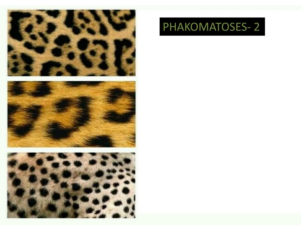
PHAKOMATOSES- 2. Z. Sturge Weber syndrome encephalotrigeminal angiomatosis. Congenital non hereditary disorder. Failure of normal development of fetal cortical veins. PORT WINE STAIN. PORT WINE = Portugal wine. PATHOLOGY.

E N D
Sturge Weber syndromeencephalotrigeminalangiomatosis • Congenital non hereditary disorder. • Failure of normal development of fetal cortical veins. PORT WINE STAIN PORT WINE = Portugal wine
PATHOLOGY • The leptomeningealhaemangioma results in a vascular steal affecting the subjacent cortex and white matter producing localisedischaemia. In about 80% of the cases, there is an unihemispheric involvement.
CTDetects subcortical calcification at an earlier age than plain film and can also demonstrate associated parenchymal volume loss • tram-track sign of cortical and subcortical calcification ,calvarial and regional sinus enlargement may be evident • ipsilateral choroid plexus may be enlarged • in severe cases, a Dyke-Davidoff-Masson appearance may be seen • MRI • T1: signal of affected region largely normal, with anatomic volume loss evident at older age • T1 C+ (Gd) • prominent leptomeningeal enhancement in affected area • much later in life the angioma may 'burn out' losing enhancement • enlarged ipsilateral choroid plexus • T2 • low signal in white matter subjacent to angioma representing • postulated accelerated myelination in neonate • calcification later in life • abnormal deep venous drainage seen as flow voids • GE/SWI/EPI: sensitive to calcification, seen as regions of signal drop out • MR spectroscopy: decreased NAA .
T1: signal of affected region largely normal, with anatomic volume loss. COR T1
T2 weighted image, showing atrophy of the right hemisphere, enlargement of the choroid plexus (arrow 1), a thickened overlying skull and subcortical calcification (hypointense / dark signal in the subcortical region - arrow 2).
T1 C+ (Gd) • prominent leptomeningeal enhancement in affected area with • enlarged • right • sided • choroid
GE/SWI/EPI: sensitive to calcification seen as regions of signal drop out
Hemimegalencephaly • Dyke davidoffmasson syndrome • Rasmussen encephalitis: tends not to have calvarial changes
Dyke-Davidoff-Masson syndrome(DDMS) was initially described as changes in the skull seen on skull x-ray in patients with cerebral hemiatrophy but is now applied more broadly to cross-sectional imaging also. It is characterised by: thickening of the skull vault (compensatory) enlargement of the frontal sinus (also ethmoidal and mastoid air-cells) elevation of the petrous ridge ipsilateralfalcine displacement capillary malformations (are a novel finding for children with Dyke-Davidoff-Masson syndrome DYKE DAVIDOFF MASSON APPEARANCE
HEMIMEGALENCEPHALY MRI of the brain demonstrates the right cerebral hemisphere to be markedly enlarged. The grey matter is disorganized and thickened, consistent with migration arrest. The right lateral ventricle demonstrates an abnormal morphology and is also markedly enlarged. There is diffuse T2 hyperintensity throughout the white matter of the right hemisphere. The right cranial vault is also expanded. Additionally the right cerebellar hemisphere is also enlarged with abnormal cortical formation. The left hemisphere is essentially normal in appearance.
RASMUSSEN’S ENCEPHALITIS ASSMUSSENS A chronic viral infection of the brain affecting only one hemisphere with unilateral cerebral hemiatrophy resulting in drug-resistant epilepsy, and progressive neurological and cognitive deterioration. Mild diffuse uniform atrophic changes of the left cerebral hemisphere manifested as deepening of cortical sulci and mildly decreased volume of cortex and white matter. This is associated with mild exvacuodilatation of the left lateral ventricle. Mild diffuse T2 and FLAIR hyperintensity is noted affecting the white matter of the left cerebral hemisphere. Mild attenuation of the left middle cerebral artery is also noted. Preserved volume and MRI features of both cerebellar hemispheres.
Von HippelLindau disease Due to mutations in the VHL tumour suppressor gene on chromosome 3.
CHOROID PLEXUS PAPILLOMA Choroid plexus papillomas are an uncommon, benign (WHO grade I) neuroepithelialintraventriculartumour which can occur in both the paediatric (more common) and adult population. On imaging, these tumours are usually identified in the fourth ventricle in adults and in the lateral ventricles in the paediatric population. They commonly present as a solid vascular tumour with vivid frond-like enhancement pattern. In a quarter of cases, speckled calcifications are present.
T1: typically isointense compared to adjacent brain; may be somewhat hypointense
T2iso to hyperintense small flow-voids may be seen within the tumour
T1 C+ (Gd): marked enhancement, tends to be homogeneous MR spectroscopy decreased NAA increased Cho
ENDOLYMPHATIC SAC TUMOR IMAGING T1 hyperintense, T2 hyperintense, multilobulated mass at the location of the vestibular aqueduct.
It gives a picture of bone erosion and the "moth-eaten" petrous bone. Endolymphatic sac tumours commonly enhance intensely on CT.
INCONTINENTIA PIGMENTI Incontinentiapigmenti, also known as Bloch-Sulzberger syndrome, is a rare condition that can affect many body systems, specially the skin. As an X-linked dominant genetic disorder, it occurs much more often in females than in males. • CNS • microcephaly • cerebral atrophy • hypoplasia of corpus callosum • periventricular white matter damage • strokes • hydrocephalus • porencephalic cysts • neuronal heterotopias
MR imaging on the 3rd day of life (A–D) and at the age of 5 months (E and F). A and B, T1-weighted images. The sagittal view (B) illustrates the extensive cortical damage including cerebellar cortex. C and D, Axial T2-weighted images, demonstrating high signal intensity in the subcortical white matter mainly of the left hemisphere and also signal intensity changes of almost the entire left cortex. On T1-weighted images, these white matter areas appear hypointense (A). E and F, At follow-up, fluid-attenuated inversion recovery images show residual cystic lesions, more on the left than on the right, involving mainly subcortical white matter and also cortical areas. There is no normal signal intensity of myelination. Basal ganglia appear atrophic on the left (F).
Hypomelanosis of ITO Skin lesions are hypopigmentedzones or spots with irregular borders, sometimes whorls, or linear white streak lines following Blaschko’slines. • CNS • Dilated VR spaces • hypoplasia or cerebellar atrophy • focal or generalized cerebral atrophy • neuronal heterotopias • hemimegalencephaly • HI has been associated rarely with vascular anomalies as Moyamoyasyndrome. The exact cause of hypomelanosis of Ito is unknown with many cases being associated with genetic mosaicism and sporadic gene mutations.
Brain MRI disclosing marked enlarged Virchow-Robin spaces in axial (A) and sagittal (B) unenhanced T1-weighted images and in coronal (C) and axial (D) enhanced T1-weighted images. Axial T2-weighted (E) and FLAIR (F) images shows enlarged Virchow-Robin spaces with mild surrounding gliosis depicted as T2 and FLAIR hyperintense foci.
GROLIN GOLTZ SYNDROME – BASAL CELL NAEVUS SYNDROME It is related to a mutation in PTCH tumor suppressor gene on chromosome 9 • CNS • Agenesis of the corpus callosum • medulloblastoma: especially in males • macrocephaly • calcified falxcerebri • calcified tentorium. • Cranio facial. • odontogenickeratocysts,often multiple • frontal bossing • hypertelorism • Macrocephaly.
Multiple radiolucent periodontogenic lesions with smooth border and cortical thinning.
AGENESIS OF CORPUS CALLOSUM Axial CT image showing dilated occipital horn of bilateral lateral ventricles
AGENESIS OF CORPUS CALLOSUM CORONAL T2W MR I IMAGE SHOWING HIGH RIDING BILATERAL FRONTAL HORNS OF LATERAL VENTRICLES
Coronal CT image showing calcification of the midline falx and tentorium
Saggital CT image showing calcification of the midline falx .