Download

1 / 22
230 likes | 373 Views
Lec. 1. أستاذ . د.رائد عزيز. PERIODONTICS. Periodontics : That branch of dentistry that deals with diagnosis and treatment of diseases and condition of the supporting and surrounding tissues of the teeth or their implanted substitutes. Periodontology :

E N D
Lec. 1 أستاذ . د.رائد عزيز PERIODONTICS
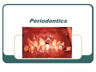
Periodontics: That branch of dentistry that deals with diagnosis and treatment of diseases and condition of the supporting and surrounding tissues of the teeth or their implanted substitutes. Periodontology: The scientific study of the periodontium in health and disease.
Periodontium comprises the following tissues: 1- The gingiva. 2- The periodontal ligament. 3- The root cementum. 4- The alveolar bone.
The gingiva: The gingiva is defined as the part of the oral mucosa that covers the alveolar processes of the jaws and surrounds the necks of the teeth. The oral mucosa consists of three zones; • The gingiva and the covering of the hard palate termed (the masticatory mucosa). • The dorsum of the tongue, covered by (specialized mucosa). • The oral mucous membrane lining the remainder of the oral cavity.
Macroscopical anatomy: In the coronal direction the coral pink gingiva terminates in the (free gingival margin) which has a scalloped outline. In the apical direction the gingiva is continuous with the loose, darker red alveolar mucosa (lining mucosa) from which the gingiva is separated by a border line called mucogingival junction.
The gingiva is divided into 2 parts:The free gingiva (FG).The attached gingiva (AG). • FG extends from (gingival margin) apically to the (free ging. groove) which is positioned at a level corresponding to the level of cementoenamel junction (CEJ).
GINGIVAL MARGIN • Margin is rounded, and forms a sulcus between the tooth and the gingiva. After complete eruption of tooth, the FG margin is located on the enamel surface approximately 0.5-2 mm coronal to the CEJ.
The attached gingiva: Extends from free gingival groove in coronal direction to mucogingival junction apically where it becomes continuous with the alveolar (lining mucosa) (AM).
Interdental papillae(I.D.P): Is that portion of gingiva that fills the interproximal space between adjacent teeth.
In anterior regions of dentition the I.D.P. is of pyramidal form while in the molar regions they are flattened in buccolingual direction. In premolar and molar regions the teeth have contact surfaces rather than contact points so the I.D.P. has a shape in conformity with the contact surfaces, so concavity is (col region) is established in the premolar and molar separating the I.D.P. in one vestibular and one lingual/palatal portion.
Microscopic anatomy: The gingival epithelium is stratified squamous epithelium which is supported by fibrous connective tissue which is called lamina properia. The epithelium covering the gingiva may be differentiated as follows:
Oral epith. which faces the oral cavity. Oral sulcularepi. which faces the tooth without touching it. Junctional epi. which provides the contact between the gingiva and thetooth surface.
The boundary between the oral epithelium (OE) and the underlying connective tissue (C.T.) has a wavy course. The C.T. portions project into the epi. are called (C.T. papillae) and are separated from each other by epithelial ridges- so- called (rete pegs).
In normal non-inflamed gingiva, rete pegs and C.T. papillae are lacking at the boundary between J.E. and its underlying C.T. So that, a characteristic feature of the O.E. and the S.E. is the presence of rete pegs, while these structures are lacking in J.E.
The oral epi. is a keratinized stratified, squamous epi. can be divided into the following: 1) Stratum basale. 2) Stratum spinosum. 3) Stratum granulosum. 4) Stratum corneum.
Other cells of OE: In addition to previously mentioned keratin producing cells which comprise about 90% of total cells there are three types of cells • Melanocytes: pigment producing cells. • Langerhans cells: play a role in defense mechanism. • Non-specific cells: form other types of cells.
The composition of desmosomes: A desmosome consists of two adjoining hemidesmosomes separated by a zone containing electron-dense granulated material (GM), and a hemidesmosome comprises the following structural components: 1) The outer leaflets (OL) of the cell membrane of two adjoining cells. 2) The thick inner leaflets (IL) of the cell membrane. 3) The attachment plaques (AP), which represent granular and fibrillar material in the cytoplasm.