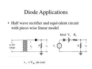
Download
1 / 4
40 likes | 79 Views
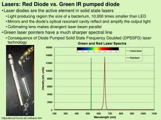
Now-a-days, almost every specialty area of dentistry is utilizing the powers of laser<br>energy for therapeutic purposes. The diode laser is the most popular soft tissue laser and<br>commonly used in dentistry. A diode laser with wavelengths ranging from 810 to 980 nm<br>in a continuous or pulsed mode is used as a possible instrument for soft tissue surgery<br>in the oral cavity.

E N D
Original Article Case Report Applications of Diode Lasers in Periodontics: A Case Series Shanmukha Srinivas Manikanta Kumar Tirumalasetty1, Gummadi Anusha1, Masapu Anupama1, Bharathi Devi Jonnalagadda1, Shyam Sunder Salavadhi2* and Kotya Naik Maloth3 1Department of Periodontics, GSL Dental College, Andhra Pradesh, India; 2Department of Periodontics, Mamata Dental College, Khammam, Telangana, India; 3Department of Oral Medicine and Radiology, Mamata Dental College, Khammam, Telangana, India Abstract Corresponding author: Dr. Shyam Sunder Salavadhi, Department of Periodontics, Mamata Dental College, Khammam, Telangana, India, Tel: 9959713715 E-mail: dr.samsunder@gmail.com Now-a-days, almost every specialty area of dentistry is utilizing the powers of laser energy for therapeutic purposes. The diode laser is the most popular soft tissue laser and commonly used in dentistry. A diode laser with wavelengths ranging from 810 to 980 nm in a continuous or pulsed mode is used as a possible instrument for soft tissue surgery in the oral cavity. The 810 nm devices predominate in dentistry, whereas 810 nm is specific for hemoglobin absorption. Diode lasers have many advantages over traditional surgical and therapeutic techniques in addition to providing bloodless surgical and post- surgical course with minimal swelling, scarring, and others things noted are less pain, excellent hemostasis, rapid wound healing and bactericidal action with the minimally invasive procedure. In the present paper, we discuss various uses of the diode laser such as esthetic crown lengthening procedure, excisional biopsy of pyogenic granuloma and gingival pigmentation in dentistry. Keywords: Diode laser; Depigmentation; Pyogenic granuloma Introduction of dark gums in both the jaws. [Figure 2a] On examination gingival pigmentation was displayed while smiling. Dummett–Gupta Oral Pigmentation Index (DOPI): Dummett [4] score was three which is moderate gingival pigmentation. Laser depigmentation was carried out for both maxillary and mandibular arches using the following settings [Figures 2b, 2c and 2d]. Laser is an acronym for “Light Amplification by Stimulated Emission of Radiation.” Laser is a Monochromatic, uni-directional and coherent beam of radiation that is produced by stimulated emission, a state where there are more excited atoms (i.e., more atoms in upper of two energy levels than in lower level), a condition called population inversion to obtain a radiation output greater than the incident radiation. [1] Many lasers with different active media were introduced since the advent of Maiman’s ruby laser. [1,2] Diode lasers are most commonly used lasers in dentistry for soft tissue procedures. The aim of this paper is to highlight the various uses of diode lasers in periodontal surgical procedures. The parameters used are: Pulsed mode – 50 µsec pulse duration with 100 µsec pulse interval Wavelength – 810 ± 10 nm (Denlase Dental Laser) Fiber diameter – 400 µm Clinical Applications of Diode Lasers Case 1: Esthetic crown lengthening Power – 4 W Mean Power – 1.34 W A 26-year-old male patient was presented with a chief complaint of gummy smile. [Figure 1a] On examination there was compromised smile esthetics because of excessive gingival display on smiling, the smile line was standard, and there is no vertical maxillary excess. The patient is systemically healthy and is not under any medication. A diagnosis of altered passive eruption type 1 sub-group A was made after periodontal examination according to Coslet et al. [3] Esthetic crown lengthening using diode lasers were done using the following settings [Figures 1b, 1c and 1d]. Case 3: Pyogenic granuloma A 29-year-old female patient reported with a chief complaint of swollen gums in the lower front tooth region, the lesion gradually increased in size throughout three months and said occasional bleeding while brushing. [Figures 3a] The patient was systemically healthy and was not using any drugs. Intraoral examination revealed a solitary, sessile lobulated gingival overgrowth extending on lingual surfaces of 42 and 43. It was reddish pink in color with white patches and was approximately 20 mm × 11 mm in size. Oval in shape with overlying surface was smooth. Radiographs like intraoral periapical radiograph showed no abnormalities, and a provisional diagnosis of pyogenic granuloma was made with a differential diagnosis of fibroma, peripheral The parameters used are: Pulsed mode – 50 µ sec pulse duration with a 100 µ sec pulse interval Wavelength – 970 ± 15 nm (Fiona Dental Laser) Fiber diameter – 400 µm This is an open access article distributed under the terms of the Creative Commons Attribution‑NonCommercial‑ShareAlike 3.0 License, which allows others to remix, tweak, and build upon the work non‑commercially, as long as the author is credited and the new creations are licensed under the identical terms. Power – 5 W Mean Power – 1.67 W Case 2: Depigmentation How to Cite this Article: Kumar MSST, et al. Applications of Diode Lasers in Periodontics: A Case Series. Ann Med Health Sci Res. 2019;9:426-429 An 18-year-old female patient was reported with a chief complaint © 2019 Annals of Medical and Health Sciences Research 426
Kumar MSST, et al.: Applications of Diode lasers in Periodontics Figure 1: (a) Gingival smile (b) Incision given with laser (c) Immediate post‑operative (d) Esthetic gingival contour. Figure 2: (a) Dummett’s class II gingival pigmentation (b) depigmentation using a laser (c) Immediate post‑operative (d) 6 months follow‑ up showing esthetic gingival color. Figure 3: (a) Pyogenic granuloma (b) Excision using the laser (c) Immediate post‑operative (d) Two weeks post‑operative. Annals of Medical and Health Sciences Research | Volume 9 | Issue 1 | January-February 2019 427
Kumar MSST, et al.: Applications of Diode lasers in Periodontics ossifying fibroma, peripheral giant cell granuloma, and hemangioma. Excision was done using a laser, and the following settings were used [Figures 3b, 3c, and 3d]. Administration (FDA) are ablating, incising, excising and coagulation for all types of intraoral soft tissue surgery. Specific approvals include; aphthous ulcer treatment, sulcular debridement, removal of coronal pulp, adjunct to root canal procedures, pulpotomy as adjunct to root canal retreatment, tooth whitening, aid in diagnosis of dental caries, blood flow measurements, treatment of herpetic lesions, coagulation of extraction sites, reduction of bacterial levels (decontamination) and inflammation, aid in detection and localization of subgingival dental calculus, and removal of highly inflamed edematous tissue affected by bacterial penetration of the pocket lining and junctional epithelium. [18,20] The parameters used are: Continuous mode Wavelength – 810 ± 10 nm (Denlase Dental Laser) Fiber diameter – 400 µm Power – 2 W Continuous wave (CW) vs. pulsed. Several studies at that time showed that continuous wave mode at 1W power was enough to reach a bactericidal effect on roots, root canals, and implant surfaces. [16,17,21] Application of 3 to 4 W in continuous modeled very fast to carbonization of the soft tissue which causes higher absorption followed by heavy thermal damage and necrosis of the tissue. To approach better results in soft tissue treatment without much carbonization, it was necessary to interrupt the CW mode. That was done by chopping the CW mode. Pulses down to several 100 µsec were utilized. Mean Power – 2 W Discussion We present three case reports that highlight the importance of laser usage in dentistry. Among various lasers, diode laser has become a crucial tool in the dental armamentarium due to its exceptional ease of use and affordability. The key advantage with diode laser is, its wavelength spectrum (810–1064 nm) is well absorbed by melanin, hemoglobin, and other chromophores that are commonly present in periodontal tissues. [4,5] With Diode Lasers the laser energy can be transmitted through a thin fiber as small as 200µ so that it can easily penetrate deep periodontal pockets to deliver its therapeutic effects. [5] Along with these features, it also has the added advantages such as portability, convenience and cost efficiency. Hence it can be easily incorporated into the dental practice. [5] Conclusion Although there is an ongoing debate over the efficiency and cost- benefit ratios of using lasers, there are substantial benefits of using a laser especially a diode laser such as reduced pain, hemostasis, and improved patient comfort. Hence lasers may be considered as a boon rather than bane for the dentistry. Diode Lasers are expected to help tissues in an inflamed and/or damaged state, enter the healing and regenerative phases by thorough debridement and decontamination of diseased tissues rapidly, and by modulating or activating cell metabolism in the surrounding tissues. [6,7] In the last decade, it has been suggested that laser irradiation alters cellular behavior by affecting the mitochondrial respiratory chain or membrane calcium channels and that it can facilitate collagen synthesis, angiogenesis, and growth factor release, which eventually accelerate wound healing. [8-12] Diode lasers have been used in the treatment of periodontal disease and have shown complete epithelial removal and irradiation of periodontal pockets has been shown to have an antimicrobial effect termed as bacterial laser reduction. This complete removal of epithelium could delay epithelial down growth and allow connective tissue attachment to occur leading to new attachment. [13] Conflict of Interest The authors disclose that they have no conflicts of interest. References 1. Hitz CB, Ewing JJ, Hecht J. Introduction to laser technology. 4th ed. Hoboken, N.J: Wiley-IEEE Press; 2012;23-25. 2. Maiman TH. Stimulated optical radiation in ruby lasers. Nature. 1960;187:493. 3. Coslet JG, Vanarsdall R, Weisgold A. Diagnosis and classifica- tion of delayed passive eruption of the dentogingival junction in the adult. Alpha Omegan 1977;70:24-28. Currently, there is minimal evidence to support the use of a laser for subgingival debridement, either as a monotherapy or adjunct to SRP. [14,15] However, there are many unique advantages of Lasers in surgical aspects of periodontal therapy such as hemostasis, reduced pain, accelerated wound healing, bactericidal action, and minimally invasive nature. [16,17] 4. Dummett CO, Barens G. Oromucosal pigmentation: An updated literary review. J Periodontol 1971;42:726-736. 5. Schwarz F, Aoki A, Sculean A, Becker J. The impact of laser application on periodontal and peri-implant wound healing. Peri- odontol 2000 2009;51:79-108. 6. Aoki A, Mizutani K, Schwarz F, Sculean A, Yukna RA, Takasaki AA, et al. Periodontal and peri-implant wound healing following laser therapy. Periodontol 2000 2015;68:217-269. Types of diode lasers The active medium of the diode laser is a solid-state semiconductor made of aluminum, gallium, arsenide, and occasionally indium; For the most part, the high power diode lasers used in laser medicine, and to pump solid-state lasers, are aluminum gallium arsenide (AIGaAs) lasers that provide a nominal laser wavelength of 800 nm, or indium gallium arsenide (InGaAs) lasers that provide wavelengths around 980 nm. [1,18] Wavelengths are ranging from approximately 400 nm to 25000 nm. In dentistry most commonly used wavelengths are 810 and 980nm. [1] The 810 nm wavelength is specific for hemoglobin absorption; the 940nm wavelength provides a balanced ratio between the light irradiated into the tissue and the absorption by hemoglobin and water; the 980nm wavelength is such that it is specific for hemoglobin and water. [19] 7. Jin JY, Lee SH, Yoon HJ. A comparative study of wound healing following incision with a scalpel, diode laser or Er, Cr: YSGG laser in guinea pig oral mucosa: A histological and immuno-histo- chemical analysis. Acta Odontol Scand 2010;68:232-238. 8. Pereira AN, De Eduardo C P, Matson E, Marques MM. Effect of low-power laser irradiation on cell growth and procollagen syn- thesis of cultured fibroblasts. Lasers Surg Med 2002;31:263-267. 9. Hawkins D, Abrahamse H. Effect of multiple exposures of low- level laser therapy on the cellular responses of wounded human skin fibroblasts. Photomed Laser Surg 2006;24:705-714. 10. Alexandratou E, Yova D, Handris P, Kletsas D, Loukas S. Human fibroblast alterations induced by low power laser irradiation at the single cell level using confocal microscopy. Photochem Photobiol Sci Off J Eur Photochem Assoc Eur Soc Photobiol 2002;1:547-552. Clinical applications Among the procedures cleared for marketing by the Food and Drug Annals of Medical and Health Sciences Research | Volume 9 | Issue 1 | January-February 2019 428
Kumar MSST, et al.: Applications of Diode lasers in Periodontics 11. Silveira PCL, Streck EL, Pinho RA. Evaluation of mitochondrial respiratory chain activity in wound healing by low-level laser therapy. J Photochem Photobiol B 2007;86:279-282. through irradiation with a diode laser: a pilot study. J Clin Laser Med Surg 1997;15:33-37. 17. Moritz A, Gutknecht N, Goharkhay K, Schoop U, Wernisch J, Sperr W. In vitro irradiation of infected root canals with a di- ode laser: results of microbiologic, infrared spectrometric, and stain penetration examinations. Quintessence Int Berl Ger 1985 1997;28:205-209. 12. Marques MM, Pereira AN, Fujihara NA, Nogueira FN, Eduardo CP. Effect of low-power laser irradiation on protein synthesis and ultrastructure of human gingival fibroblasts. Lasers Surg Med 2004;34:260-265. 13. Romanos GE, Henze M, Banihashemi S, Parsanejad HR, Winck- ler J, Nentwig G-H. Removal of epithelium in periodontal pockets following diode (980 nm) laser application in the animal model: An in vitro study. Photomed Laser Surg 2004;22:177-183. 18. Hilgers JJ, Tracey SG. Clinical uses of diode lasers in orthodon- tics. J Clin Orthod JCO 2004;38:266-273. 19. Yu DY, Chen HC, Chang SY, Hsiao YC, Chang CJ. Comparing the Effectiveness of 1064 vs. 810 nm Wavelength Endovascular Laser for Chronic Venous Insufficiency (Varicose Veins). Laser Ther 2013;22:247-253. 14. American Academy of Periodontology Statement on the Efficacy of Lasers in the Non-Surgical Treatment of Inflammatory Peri- odontal Disease. J Periodontol 2011;82:513-514. 20. Graeber JJ. Diode Lasers: A Primer. www.dentalacademyofce. com/courses/2564/PDF/1401ceiGraeber_rev3.pdf. 15. Statement on Lasers in Dentistry 2018. Available from: https:// www.ada.org/en/about-the-ada/ada-positions-policies-and-state- ments/statement-on-lasers-in-dentistry 21. Gutknecht N, Van Gogswaardt D, Conrads G, Apel C, Schubert C, Lampert F. Diode laser radiation and its bactericidal effect in root canal wall dentin. J Clin Laser Med Surg 2000;18:57-60. 16. Moritz A, Gutknecht N, Doertbudak O, Goharkhay K, Schoop U, Schauer P, et al. Bacterial reduction in periodontal pockets Annals of Medical and Health Sciences Research | Volume 9 | Issue 1 | January-February 2019 429