Download
1 / 34
340 likes | 356 Views
GRAND ROUNDS Anterior Segment OCT Imaging in a c ase of Acute Anterior Uveitis. Mehreen Adhi, MD October 21, 2016. Patient Presentation. Chief Complaint: “My left eye hurts really bad and I cannot see anything out of it” HPI:

E N D
GRAND ROUNDSAnterior Segment OCT Imaging in a case of Acute Anterior Uveitis Mehreen Adhi, MD October 21, 2016
Patient Presentation Chief Complaint: • “My left eye hurts really bad and I cannot see anything out of it” HPI: • 55 year old diabetic white gentleman presented to the retina clinic in August 2016 at the Robley Rex VA Medical Center with pain, photophobia and blurring of vision OS for 5 days. • He was vacationing in Las Vegas when symptoms came on suddenly • He had Cyclopentolate from one of his previous flare ups that he started using before presenting • Per patient, his left eye had “flared up” at least 3 times in the past
Patient Presentation HPI (continued…): • First episode: June 2013 – resolved with topical steroids • Second episode: March 2014 – resolved with topical steroids - systemic work up done at this time • Third episode: December 2015 – resolved with topical steroids
Patient Presentation Review of Systems: • General: no fever, fatigue, weight loss • Cardiovascular: unremarkable • Respiratory: no flu-like symptoms, sinusitis, hemoptysis, shortness of breath • Gastrointestinal: h/o chronic diarrhea • Genitourinary: unremarkable • Neurological: unremarkable • Musculoskeletal: h/o intermittent back pain • Integumentary: no rash or skin lesions
Patient Presentation Review of Systems: • General: no fever, fatigue, weight loss • Cardiovascular: unremarkable • Respiratory: no flu-like symptoms, sinusitis, hemoptysis, shortness of breath • Gastrointestinal: h/o chronic diarrhea • Genitourinary: unremarkable • Neurological: unremarkable • Musculoskeletal: h/o intermittent back pain • Integumentary: no rash or skin lesions
Patient Presentation Past Ocular History: • No h/o trauma to either eye; no h/o similar episodes in OD • Mild non-proliferative diabetic retinopathy OU • Glaucoma suspect OU: based on cup/disc ratio • Nuclear sclerotic cataract OU Past Medical History: • Diabetes (insulin dependent) Past Surgical History / Family History: • Unremarkable Social History: • Former smoker; occasional/social alcohol use; no recreational drugs Medications: • Long-acting insulin (Glargine) and pre-prandial insulin sliding scale Allergies: • No known drug allergies
Posterior Segment Exam B-scan: Vitreous clear; Retina flat
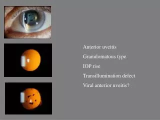
Clinical Photos OD OS
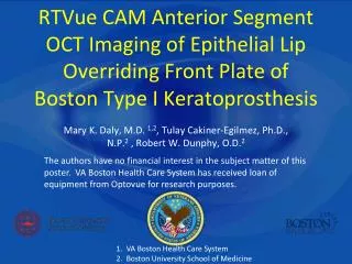
Anterior Segment OCT 386 um 282 um
Assessment • 55 year old diabetic white gentleman with a 5 day history of blurred vision, photophobia and pain in the left eye; exam significant for 2-3+ anterior chamber flare with a 360 degrees fibrin membrane overlying the anterior aspect of the lens OS • Recurrent Acute Non-granulomatous Acute Uveitis OS
Plan and Follow up • Pred acetate ophthalmic solution Q1H OS • Medrol dose pack PO • Cyclopentolate ophthalmic solution TID OS • Alphaganophthalmic solution BID OS • Follow up 2 days later: sub-tenonTriescence OS • Follow up 3 weeks later….
Clinical Photos OD OS
Discussion • Anterior segment optical coherence tomography (AS-OCT) allows the visualization of various features of the anterior segment • In-vivo cross-sectional imaging of the anterior segment from AS-OCT is particularly useful in the presence of corneal opacity and ocular inflammation • Non-invasive ancillary test for assessment of features of anterior uveitis, its complications, and response to treatment
Discussion • Corneal thickness/edema Healthy subject Acute anterior uveitis
Discussion • Corneal thickness/edema Healthy subject Acute anterior uveitis
Discussion Anterior segment optical coherence tomography in acute anterior uveitis Cristiana Agra, Lydianne Agra, Jeanine Dantas, Tiago EugênioFaria e Arantes, JoãoLins de Andrade Neto Arq. Bras. Oftalmol. Feb 2014; 77:1
Discussion • Keratic precipitates
Discussion • Fibrin membrane
Discussion • Inflammatory cells in the anterior chamber
Discussion High-speed optical coherence tomography for imaging anterior chamber inflammatory reaction in uveitis: clinical correlation and grading. Agarwal A, Ashokkumar D, Jacob S, et al Am J Ophthalmol 2009 Mar;147(3):413-416.e3.
Discussion Automated Analysis of Anterior Chamber Inflammation by Spectral-Domain Optical Coherence Tomography. Sharma S, LowderCY, Baynes K, et al Ophthalmology 2015 Jul;122(7):1464-70
Conclusions • Anterior segment optical coherence tomography (AS-OCT) may be a useful non-invasive ancillary test in patients with anterior uveitis • Features such as corneal thickness/edema, keratic precipitates, fibrin deposition and anterior chamber inflammation may be useful parameters to assess treatment response
Acknowledgements • Shorye Payne MD • Mary and Tammy • Drs. Syed, Fernandez, Kassm, Breaux, Piri, Mueller
References • Cristiana Agra, Lydianne Agra, Jeanine Dantas, Tiago EugênioFaria e Arantes, JoãoLins de Andrade Neto. Anterior segment optical coherence tomography in acute anterior uveitis. Arq. Bras. Oftalmol. Feb 2014; 77:1 • Agarwal A, Ashokkumar D, Jacob S, et al. High-speed optical coherence tomography for imaging anterior chamber inflammatory reaction in uveitis: clinical correlation and grading. Am J Ophthalmol. 2009;147:413–416. e413. • Sharma S, Lowder CY, Vasanji A, Baynes K, Kaiser PK, et al. Automated Analysis of Anterior Chamber Inflammation by Spectral-Domain Optical Coherence Tomography. Ophthalmology 2015 Jul;122(7):1464-70. • Regatieri CV, Alwassia A, Zhang JY, et al. Use of Optical Coherence Tomography in the Diagnosis and Management of Uveitis. IntOphthalmolClin 2012 Fall; 52(4): 33-34 • Lowder CY, Li Y, Perez VL, DH Anterior Chamber Cell Grading with High-Speed Optical Coherence Tomography. Invest Ophthalmol Vis Sci. 2004;45 E-Abstract 3372.