Download
1 / 10
420 likes | 2.17k Views
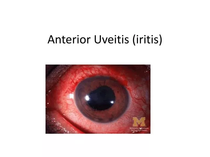
Anterior Uveitis ( iritis ). Anatomy. U veitis = inflammation of the uvea Uvea = iris, cilliary body, choroid Anterior uveitis = inflammation of iris and anterior chamber

E N D
Uveitis = inflammation of the uvea • Uvea = iris, cilliary body, choroid • Anterior uveitis = inflammation of iris and anterior chamber • Intermediate uveitis= inflammation of middle part of the uvealtract, mainly the vitreous humour. It can also affect the underlying retina. • Posterior uveitis= inflammation which affects the back (posterior) part of the eye. It can affect the choroid, the head of the optic nerve, and the retina (or any combination of these structures). It includes chorioretinitis, retinitis and neuroretinitis. • Panuveitis = inflammation affecting the whole of the uvealtract
WHO ? • Most common in young/middle aged adults 20-59yrs • Affects 17-52 people per 100,000 per yr Causes ? • 50 % are idiopathic • 50 % of those with anterior uveitis are HBLA27 +VE • May be secondary to corneal graft • Eye infections e.g. toxoplasmosis, herpes virus keratitis
Other associations .. • Autoimmune : HLAB27, Reiters syndrome, Bechets • Infectious : leptospirosis, lyme disease, syphilis,TB • Systemic disease : IBD, MS, kawsaki’s, juvenile arthritis, polyarteritisnodosa,psoriatic/reactive arthritis, sarcoidosis, SLE • Drugs : Rifabutin ( similar to rifampicin) • Trauma • Cancer : NHL, HL, Leukaemia, Melanoma
Presents .. Symptoms • Acute onset pain • Photophobia • Blurred vision • Decreased visual acuity • Headache
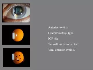
Signs • Watering • Circumcorneal redness • Small or irregular pupil • +/- hypophyon ( anterior chamber pus causing white ‘fluid-level’ line)
+/- keratitic precipitates on posterior surface of cornea • Cells in anterior chamber on slit lamp exam • Increasing pain as the eyes converge and the pupils constrict
Management • Urgent opthalmology referral (within 24hrs) normally topical/oral glucocorticoidsteriods • Cycloplegic drops for comfort e.g. Atropine • Simple analgesia
Complications • Cystoid macular oedema • Secondary cataract • Posterior synechiae (irregular pupil shape) • Raised IOP Glaucoma, secondary to either inflammatory process or steriods • Vitreous opacities • Retinal detachment • Neovascularisation of the retina/optic nerve/iris • Relapses are common BUT with prompt and effective treatment 91% return to normal vision