Download
1 / 12
130 likes | 377 Views
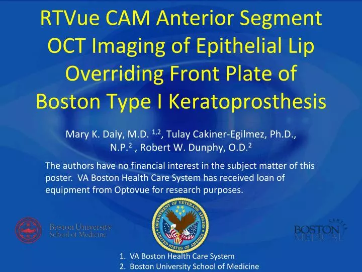
RTVue CAM Anterior Segment OCT Imaging of Epithelial Lip Overriding Front Plate of Boston Type I Keratoprosthesis. Mary K. Daly, M.D. 1,2 , Tulay Cakiner-Egilmez, Ph.D., N.P. 2 , Robert W. Dunphy, O.D. 2.

E N D
RTVue CAM Anterior Segment OCT Imaging of Epithelial Lip Overriding Front Plate of Boston Type I Keratoprosthesis Mary K. Daly, M.D. 1,2, Tulay Cakiner-Egilmez, Ph.D., N.P.2 , Robert W. Dunphy, O.D.2 The authors have no financial interest in the subject matter of this poster. VA Boston Health Care System has received loan of equipment from Optovue for research purposes. 1. VA Boston Health Care System 2. Boston University School of Medicine
Purpose: Endophthalmitis is a major concern following keratoprosthesis (KPro) surgery and one of the most devestating sequelae. Integration of any KPro with recipient tissue is crucial to avoiding extrusion and to ensure a sealed, closed system. The Boston Type I KPro is comprised of a PMMA front plate and stem, a backplate, and a locking ring. These components are assembled with donor cadaver corneal tissue between the front and back plate. The donor corneal skirt of the assembled KPro is then sutured into the recipient corneal bed. Full time soft contact lens (BCL) wear is recommended post-operatively to protect the front plate and improve hydration and viability of the corneal tissue around the KPro. Visualization of corneal epithelial growth around/over the edge of the front plate would be a reassuring sign for surgeons that a barrier to microbial ingress exists.Methods: Optovue RTVue CAM Anterior Segment OCT images of 4 eyes of 3 patients from the VA Boston Healthcare System with Boston Type I KPro were retrospectively reviewed to assess relationship between the KPro PMMA front plate and donor corneal tissue.Results: OCT Images of all 4 eyes demonstrated corneal epithelium extending from the donor corneal tissue and overriding the edge of the KPro front plate, a finding which was not always evident by slit-lamp examination. One of these patients imaged in the immediate postoperative period demonstrated such growth of epithelium at one week postop.
AGE (CURRENT): 84 • OPERATED EYE: OS, 10/2007 • PREOPERATIVE DIAGNOSIS: • herpes zoster keratitis left eye, • corneal scarring, corneal vascularization • KPROTYPE: Pseudophakic Boston Type 1 Keratoprosthesis (PMMA Backplate) • CURRENT TOPICAL MEDICATIONS (OS): • 1. Vancomycin 14mg/ml OS every other day • 2. Vigamox QD • 3. Prednisolone Acetate QD • 4. Brimonidine BID • 5. Travatan QHS • 6. Refresh tears prn Patient 1Imaged without BCL Front Plate Vertical Section Front Plate Donor Cornea Skirt Epithelium Stem Recipient / Donor Junction
Epithelium Superior Front Plate Horizontal OCT sections demonstrating continuous epithelial growth over peripheral edges of front plate 2 years postop Epithelium Front Plate Patient 1 Backplate Inferior
Patient 2 OD PMMA Backplate • AGE (CURRENT):88 • OPERATED EYE: OU • SURGERY DATE: OD: 4/2004 OS: 2/2005 • KPROTYPE: OD: PMMA OS: Titanium • CURRENT TOPICAL MEDICATIONS: • 1- Vigamox OU QHS • 2- Vanco 14mg/ml OU QHS • 3- Durezol OD BID • 4- Pred Forte OS QHS • 5 -Brimonidine OU TID • 6- Trusopt OU BID OS TITANIUM Backplate
Patient 2 OD PMMA Front Plate BCL Stem Epithelium Image below modified to enhance bandage contact lens (BCL) and epithelium signal BCL Epithelium
BCL OD Patient 2 Epithelium FRONT PLATE Stem Vertical section Epithelium FRONT PLATE Horizontal section edge of front plate
OSPatient 2 HORIZ OCT INTERSECT 3:30 Note gap HORIZ OCT INTERSECT 2:30 OBLIQUE OCT INTERSECT 7:30
Patient 3 • PREOPERATIVE DIAGNOSIS OS: Failed DSEK/pseudophakic bullous keratopathy • SURGERY DATE: OS 2/26/2010 • KPRO TYPE OS: Boston Type I keratoprosthesis (TITANIUM Backplate) • POST OP DAY 1 TOPICAL MEDICATIONS OS: • 1-Vanco 14mg/ml QID • 2-Cosopt BID • 3-Travatan QHS • 4-Brimonidine TID • 5-Vigamox QID • 6-Prednisolone Acetate QID • AGE: (CURRENT) 88 • PREOPERATIVE DIAGNOSIS OD:Pseudopahkic bullous keratopathy, Fuchs, history of failed corneal grafts x 2 secondary to immune graft rejection • SURGERY DATE: OD 8/2007 • KPRO TYPE OD: Boston Type I keratoprosthesis (PMMA Backplate) • CURRENT TOPICAL MEDICATIONS OD: • 1-Vanco 14mg/ml QD • 2-Cosopt BID • 3-Travatan QHS • 4-Brimonidine TID • 5-Vigamox QID • 6-Prednisolone Acetate QID OD OS Postop Day 1
Epithelium at edge and tracking under front plate BCL Patient 3 OD Front Plate Donor Cornea Skirt Stem Recipient Cornea Backplate
POST OP Week 1 OS Patient 3 POST OP Day 1 OS Patient 3 Inferotemp Increased signal suggesting epithelium epithelium FRONT PLATE FRONT PLATE GAP Increased signal suggesting epithelium FRONT PLATE epithelium FRONT PLATE FRONT PLATE Nasal epithelium Increased signal suggesting epithelium FRONT PLATE FRONT PLATE Donor Inferior Recipient
Conclusions Conclusions: OCT is useful in demonstrating presence of an epithelial lip extending from the donor corneal rim over the edge of the Boston Type I KPro frontplate. The presence of such epithelial growth is reassuring as it suggests a tight seal and a barrier to infection.