Download
1 / 49
490 likes | 497 Views
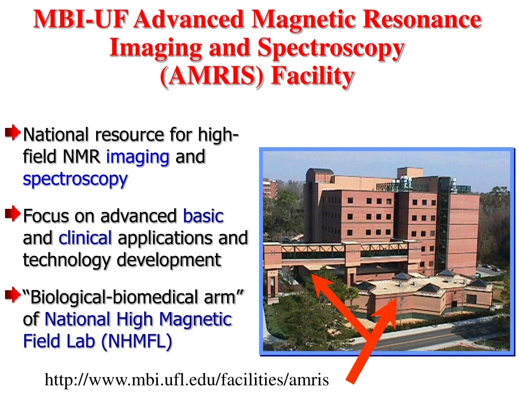
MBI-UF Advanced Magnetic Resonance Imaging and Spectroscopy (AMRIS) Facility. National resource for high-field NMR imaging and spectroscopy Focus on advanced basic and clinical applications and technology development

E N D
MBI-UF Advanced Magnetic Resonance Imaging and Spectroscopy (AMRIS) Facility • National resource for high-field NMR imaging and spectroscopy • Focus on advanced basic and clinical applications and technology development • “Biological-biomedical arm” of National High Magnetic Field Lab (NHMFL) http://www.mbi.ufl.edu/facilities/amris
MBI-UF AMRIS Instrumentation • 136 Mhz, 3.0 Tesla, 60 cm horizontal bore • MRI/S of live animals (humans, primates, dogs, etc.) • 136 Mhz, 3.0 tesla, 80 ( 94) cm horizontal bore • MRI/S of live humans • 200 Mhz, 4.7 tesla, 33 cm horizontal bore • MRI/S of live animals (cats, rabbits, rats, mice, etc.) • 473 Mhz, 11.1 tesla, 40 cm horizontal bore • MRI/S of live animals (primates, cats, rabbits, rats, mice, etc.) • Solution state NMR spectroscopy of biomolecules (multiple samples) • 500 Mhz, 11.7 tesla, 5.2 cm vertical bore • Solution/solid state NMR spectroscopy of biomolecules • 600 Mhz, 14.1 tesla, 5.2 cm vertical bore • Solution state NMR spectroscopy of biomolecules • Cryoprobe to boost S/N by a factor of 4 • MRI/S of superfused cells/tissues • 750 Mhz, 17.6 tesla, 8.9 cm vertical bore • MRI/S of superfused cells/tissues & of live animals (e.g., mice) • Solution/solid state NMR spectroscopy of biomolecules (multiple samples) • Cryoprobe under development
MBI-UF AMRIS: From Molecules to Man Single cell MRI/NMR High-Resolution Structural Biology Microsample (1.5ml) spectroscopy MR Microscopy (ex vivo) Animal MRI/MRS Human research
c o i l C t MBI-UF AMRIS RF Engineering Lab Microcoils and arrays (MRI & MRS/NMR) Superconducting probes Phased array coils Human coils Large volume/High frequency Beck et al. (2002) MAGMA 13: 152-157
MBI-UF AMRIS: 2002 User Research Highlights Brian Shilton (Univ of Western Ontario), Hargrave, Smith, McDowell, and Edison, “High-field structural studies of Rhodopsin/Arrestin complexes” Elisar Barbar (Ohio University) and Edison, “Structural biology of microtubule transport” Cottrell (St. Andrews), Zachariah, Dossey, Edison, “3D structure of a neuropeptide bound to its receptor” Webb (Illinois), Thelwall, Grant, Blackband, “NMR Microscopy of a Single Neuron Isolated from Aplysia Californica” Grant, Plant, Mareci, Blackband, Webb (Univ. Illinois), Aken (Univ. Arizona), “Proton Spectra from a Single Neuron Isolated from Aplysia Californica” Benveniste (Brookhaven Nat. Lab), Zhang (Brookhaven), Grant, Blackband, “MR Microimaging Studies of Mouse Brains For Generation of a Web Based Atlas and Methods for Identification of Brain Structures” Silver, Plant, Blackband, Benveniste (Brookhaven Nat. Lab), “Normal Mouse Brain MRI In Situ” Webb (Illinois), Zhang (Illinois), Edison, “Double Protein NMR coil”
High Field MR Technology Development Yu LI McKnight Brain Institute Advance Magnetic Resonance Imaging and Spectroscopy Facilities University of Florida, Gainesville, FL 32610
Outline • Research Background • Basic MR Principles • Small-Volume Protein NMR • MR Parameters Estimation • Imaging Technology • Summary
Roadmap • Research Background • Basic MR Principles • Small-Volume Protein NMR • MR Parameters Estimation • Imaging Technology • Summary
MR Research Areas • MR Spectroscopy • Solution state • Solid state • MR Imaging • Human/Animal imaging • Microimaging • Material imaging • Data Processing • Spectral data processing • Image reconstruction • Image post-processing
High Field MR Technology • NIH Resource • Resource Cores: • High Field Small Animal Imaging • Microimaging and Microspectroscopy • High-sensitivity and High-throughput Solution State NMR
Roadmap • Research Background • Basic MR Principles • Small-Volume Protein NMR • MR Parameters Estimation • Imaging Technology • Summary
MR Phenomena: Resonance B0 B0 Michael Sattler EMBL Heidelberg, Biomolecular NMR Structure, http://www.EMBL-Heidelberg.de/nmr/
MR Phenomena: Free Relaxation z M Mz y Mxy x y x
MR Application Fourier Transform Image Reconstruction Michael Sattler EMBL Heidelberg, Biomolecular NMR Structure, http://www.EMBL-Heidelberg.de/nmr/
MR Instrumentation Magnet RF coil and Object Gradient coil Gradient coil Receiver Transmitter ADC Synthesizer Console
Advantage: Information Rich • Molecule structure • Anatomical structure • Physiological mechanism • Pathophysiologies • Biological functional structure
Drawback: Low SNR • Spectroscopy • Low sample efficiency • Low throughput • Imaging • Long imaging time • Low resolution High Field Technology
Roadmap • Research Background • Basic MR Principles • Small-Volume Protein NMR • MR Parameters Estimation • Imaging Technology • Summary
Protein Structure Chain structure Amino Acid Primary Secondary Tertiary Quaternary
Protein NMR Michael Sattler EMBL Heidelberg, Biomolecular NMR Structure, http://www.EMBL-Heidelberg.de/nmr/
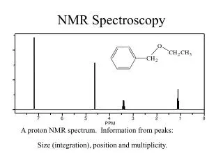
Structure Information Frequency shift • Frequency shift: chemical structure dependence • Spectral peak structure: connection between different chemical groups
Small Volume / High Field • Significance of small volume • Time of sample preparation • Expense • Availability • High field rationale B0 field RF coil design D.I.Hoult and R.E.Richards, J.Magn.Reson, 24, 71-85 (1976)
Current Probe Technology Required sample volume: 600 µL
Saddle and Solenoid Saddle Solenoid B1 Current: i Current: i “the disappointing signal-to-noise ratio experienced with superconducting system is a direct consequence of the use of saddle-shaped coils” D.I.Hoult and R.E.Richards, J.Magn.Reson, 24, 71-85 (1976)
Solenoid Coil L2 C6 C5 C1 L4 C8 C4 L3 L1 C7 C2 C3 C12 C13 C10 C9 L5 1H C11 15N 13C C15 C14 Lock Solenoid Probe Design
Roadmap • Research Background • Basic MR Principles • Small-Volume Protein NMR • MR Parameters Estimation • Imaging Technology • Summary
MR Parameters in Frequency Domain Fourier Transform Intensity Linewidth Frequency
CE NMR SNR = 22.0 SNR = 36.6 Se(f) Seb(f) B(f) Noise
Problem Formulation Srb(f) Sr(f) B(f) Noise Know Sr(f), Detect Srb(f), Estimate B(f) Seb(f) Se(f) B(f) Noise Know B(f), Detect Seb(f), Estimate Se(f)
Gradient Decent Method es(f) eb(f) _ Se(f) _ B(f) B(f) Sr(f) + + Seb(f) Srb(f) (•)2 (•)2
Gradient Decent Method Error function Parameters Optimum Values
Multiresolution detection with wavelet High resolution / High SNR Low resolution / Low SNR Wavelet transform Scale decrease S. Mallat, and W.L. Hwang, IEEE Trans. on Information Theory, Vol. 38(2), 617-643 (1992).
Resolving Results 100 mM sucrose in D2O
Roadmap • Research Background • Basic MR Principles • Small-Volume Protein NMR • MR Parameters Estimation • Imaging Technology • Summary
Image Contrast: MR Parameters-weighted • Proton density • Physical composition • T1 • Soft tissue • T2 • Tissue structure • Tissue metabolism • Pathophysiologies
Image Contrast: MR Parameters-weighted • T2* • Vascular physiology • Biological functions • Apparent Diffusion Coefficient (ADC) • Tissue microstructure • Tissue composition • Tissue constitutes • Architectural organization
3D Brain / Spinal Cord Imaging T2-weighted Images of rat brain and spinal cord High resolution: below 40 µm (17.6T) B. Beck, D.H. Plant, S.C. Grant, PlE. Thelwall, X. Silver, T.H. Mareci, H. Benveniste, M. Smith, S. Crozier, S.J. Blackband
Brain Slice Imaging Diffusion weighted microimage of rat brain slice High Resolution: 20 µm (14.1 T) S.J. Balckband, J.D. Bui, D.L. Buckley, T. Zelles, H.D. Plant, B.A. Inglis, M.I. Phillips
Neuron Cell Imaging Diffusion-weighted images of a single neuron cell Cytoplasm (C) and nuclear (N) in artificial sea water (S). High Resolution: 20 µm (14.1 T) S.C. Grant, D.L. Buckley, S. Gibbs, A.G. Webb, and S.J. Balckband
Roadmap • Research Background • Basic MR Principles • Small-Volume Protein NMR • Spectral Resolution Restoration • Imaging Technology • Summary
High Field MR Technology • Hardware development • Magnet • Coil geometry / dimension • RF circuit design • Algorithm development • MR parameters estimation • Biomedical information and MR parameters • Image processing • EM field calculation
Acknowledgement Jim Roca Paul Moliter William Brey Feng Lin Peter Gor’kov Jim Norcross Terry Green Drs Arthur Edison Andrew Webb Stephen Blackband Samuel Grant