Download
1 / 19
210 likes | 439 Views
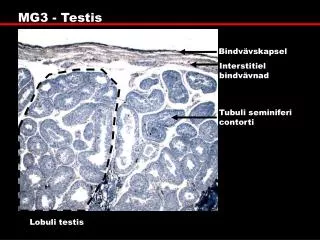
Cells of the testis. Spermatogonia Leydig (interstycial) cells: Influence of LH testosterone Sertoli cells: Optimális medium for the differentiation of a spermiums FSH, LH stimulate spermatogenézist The maturing germ cells and spermatids (left arrow)

E N D
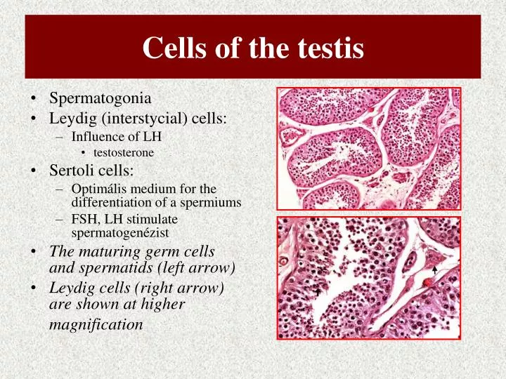
Cells of the testis • Spermatogonia • Leydig (interstycial) cells: • Influence of LH • testosterone • Sertoli cells: • Optimális medium for the differentiation of a spermiums • FSH, LH stimulate spermatogenézist • The maturing germ cells and spermatids (left arrow) • Leydig cells (right arrow) are shown at higher magnification
Biosynthsis of testosteron
Mechanism of the action of testosteron androgen receptor binds testosterone directly or its active metabolite 5a-dihydrotestosterone (DHT). dissociation of heat shock proteins (hsp) the receptor enters the nucleus steroid hormone binding either in the cytoplasm or in the nucleus, the androgen receptor binds as dimers to specific DNA elements present as enhancers in upstream promoter sequences of androgen target genes. RNA-polymerase II [RNA-Pol II], TATA box binding protein [TBP], TBP associating factors [TAF's], general transcription factors [GTF's]) triggers mRNA synthesis consequently protein synthesis results in an androgen response.
Hormons of testis • 50-100 mg DHT/day • Periferial testosteron + DHT conversion • 17b-estradiol (E2) • Leydig-, Sertoli-cells • FSH regulation? • Testosteron: • dominant: prenatál period, puberty • 5 mg/day adult men • After birth: only androsterone
[testosteron] is lowered by the years • Sex Hormon Binding Protein (SHBP) vagy (Testosterone-Estrogen Binding Protein (TEBP) • 97-99 % testosteron / SHBP + albumin • SHBP • liver • Estrogen elevates its level • testoszterone decreases
Testosteron + DHT • Sexual differenciátion • Spermatogenesis • Secundary sexual characters • Anabolic effects, gén-regulation • Benign hypertrophy of prostatse • – 5a hydroxylase inhibitiors • Lack of Testosteron biosyntesis • hipogonadizmus • 5a hydroxilase defective receptors
The role of testosteron / DHT receptor • Testicular feminization syndrome Note the absence of lumens and presence of only sertoli cells in the seminiferous tubules. Numerous Leydig cells are seen. Leydig cells in testicular feminization syndrome often lack Reinke’s crystalloids
Testosteron metabolism • Oxydation at C17 position • Reduction at C3 , double bond
Ovarium: steroid hormon synthesis • Theca interna • Stratum granulosum
Folliculus • estrogen • the endometrium to become thicker and more richly supplied with blood vessels and glands • rising level of LH causes the developing egg within the follicle to complete the first meiotic division (meiosis I), forming a secondary oocyte. • ~ 2 weeks, there is a sudden surge in the production of LH. • LH triggers ovulation • the release of the secondary oocyte into the fallopian tube. • Under the continued influence of LH, the now-empty follicle develops into a corpus luteum • Stimulated by LH, the corpus luteum secretes progesterone which • continues the preparation of the endometrium for a possible pregnancy • inhibits the contraction of the uterus • inhibits the development of a new follicle • If fertilization does not occur (which is usually the case), • the rising level of progesterone inhibits the release of GnRH which, in turn, inhibits further production of progesterone. • As the progesterone level drops, • the corpus luteum begins to degenerate; • the endometrium begins to break down, its cells committing programmed cell death (apoptosis); • the inhibition of uterine contraction is lifted, and • the bleeding and cramps of menstruation begin.
Synthetic agonists, antagonists influence the conception and tumor proliferation
Clomifene (INN) or clomiphene (USAN and former BAN) or Clomid or Clomifert is a selective estrogen receptor modulator (SERM), used mainly in female infertility due to anovulation (e.g. due to polycystic ovary syndrome). In some countries, it is also registered for use in men. Clomiphene citrate is marketed under various trade names including Clomid, Serophene, Milophene, etc. Clomifene acts by inhibiting the action of estrogen on the gonadotrope cells in the anterior pituitary gland. In response to low estrogen levels, follicle-stimulating hormone (FSH) release is increased, leading to a higher rate of ovulation and hence pregnancy. Clomifene can lead to multiple ovulation, and hence increasing the chance of twins. In comparison to purified FSH, the rate of ovarian hyperstimulation syndrome is low. There may be an increased risk of ovarian cancer and weight gain.