Download
1 / 21
210 likes | 463 Views
Géométrie. d’après G. Herzberg. d’après G. Herzberg. Variance “normale ”. +2 mm. d’après J. Laulan. L’analyse radiographique se fait sur les clichés standards face et profil. Complétés par des clichés en 3/4 à la recherche du fragment postéro-médial.

E N D
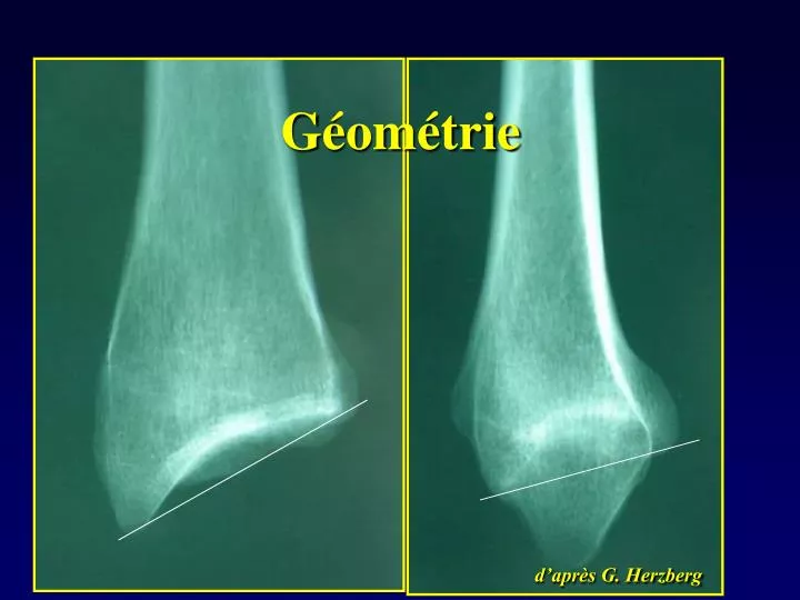
Géométrie d’après G. Herzberg
Variance “normale” +2 mm
d’après J. Laulan L’analyse radiographique se fait sur les clichés standards face et profil Complétés par des clichés en 3/4 à la recherche du fragment postéro-médial
… Et des clichés sous traction • Réalisés au bloc • Contributifs 2 fois sur 3 • Pour analyser la fracture …
Questions … 20%des fractures ! • Fréquence ? • 3 Tableaux cliniques ? • Quel est le risque si ostéoporose ? • 3 complications ? Femme 60 basse Energie - Ostéoporose Homme 50 éthylique < 40 haute énergie - motard Sur risque de fracture : 30% Déplacement II - raideur - algodystrophie
Quelles sont les causes d’une hémarthrose post traumatique du genou ? • Fractures fémur – tibia – rotule • LCA • Ménisques
LCA LCA normal Rupture du LCA en IRM
Mise en évidence d’une laxité ligamentaire Tiroir antérieur Trillat-lachman test En flexion "LACHMAN test" introduit par TORG 1976 pratiqué en France par TRILLAT (1963)
Le plus souvent, il y a une fissure postérieure qui peut évoluer Vers l’avant : Anse de seau Anse de seau bloquée Vers l’arrière : Languette post
Blocage du ménisque interne Anse de seau L’anse de seau vient s’interposer en avant du condyle
Examen clinique Point méniscal interne Grinding test (Appley)
Problèmes pratiques • Prothèse articulaire + douleur = … • Infection • Algodystrophie ( syd régional douloureux complexe de type 1) • Luxation • Descellement ( années) • Fracture … • Migration ( stent, chambre implantables)
Collapsus cardio-vasculaire Syndrome de confusion mentale Pétéchies sur le corps Signes au FO
Fractures ouvertes classification de cauchoix Stade 1 Plaie simple suturable sans tension après parage Stade 2 Plaie SUTURABLE EN TENSION APRES PARAGE Stade 3 PERTE DE SUBSTANCE