Download

1 / 23
240 likes | 267 Views
CELL MEMBRANES and TRANSPORT. dr shabeel pn. The Cell. Learning Objectives. Describe the fluid mosaic model of membrane structure and explain the underlying reasons for this structure. Outline the roles of phospholipids, cholesterol, glycolipids, proteins and glycoproteins in membranes.

E N D
CELL MEMBRANESandTRANSPORT dr shabeel pn
Learning Objectives • Describe the fluid mosaic model of membrane structure and explain the underlying reasons for this structure. • Outline the roles of phospholipids, cholesterol, glycolipids, proteins and glycoproteins in membranes. • Outline the roles of the plasma membrane, and the roles of membranes within cells. • Describe and explain how molecules can get in and out of cells (cross cell membranes) by the processes of diffusion, facilitated diffusion, osmosis, active transport, endocytosis and exocytosis. • Describe the effects on animal and plant cells of immersion in solutions of different water potential. • Describe the features of the gaseous exchange surface of mammalian lung. • Describe the features of root hairs that enable the uptake of ions by active transport.
Key words you should know • Phospholipids Solution Pinocytosis • Polar Solute Micropinocytosis • Hydrophilic Solvent Exocytosis • Hydrophobic Partially permeable Gaseous exchange • Micelles Water potential Alveoli • Phospholipid bilayer Solute Potential Root hair • Fluid mosaic model Pressure Potential Surface area • Glycoproteins Turgid Epidermis • Glycolipids Plasmolysis Passive transport • Cholesterol Plasmolysed • Proteins Incipient plasmolysis • Transport proteins Active transport • Enzymes Carrier protein • Receptor molecules Bulk transport • Diffusion Endocytosis • Concentration gradient Phagocytosis • Facilitated diffusion Phagocytes • Osmosis Phagocytic vacuoles
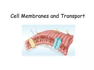
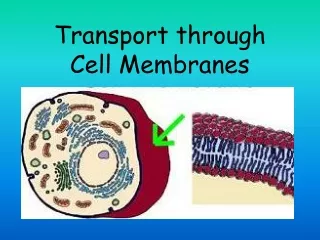
Cell membrane • All living things are surrounded by a membrane. • A cell membrane is also known as plasma membrane. • Controls exchange of materials such as nutrients and waste between cells and their environment. • Has other important functions for example to enable cells to receive hormones. • To understand the function of anything in biology, you must study the structure first!
Cell Membranes from Opposing Neurons (TEM x436,740). Nerve cell Gap between cells Cell membrane { } cell membrane 7nm wide Nerve cell
HYDROPHILIC heads (water liking)-Attracted to the water called POLAR HYDROPHOBIC tails (water fearing)-Not attracted to the water called NON-POLAR Cell membranes are made of PHOSPHOLIPIDs A Phospholipid
Phospholipids are important structural components of cell membranes. Phospholipids are modified so that a phosphate group (PO4-) replaces one of the three fatty acids normally found on a lipid. The addition of this group makes a polar "head" and two nonpolar "tails".
HYDROPHILIC HEAD At the other end of the phospholipid is a phosphate group and several double bonded oxygens. The atoms at this end of the molecule are not shared equally. This end of the molecule has a charge and is attracted to water. It is POLAR HYDROPHOBIC TAILS The two long chains coming off of the bottom of this molecule are made up of carbon and hydrogen. Because both of these elements share their electrons evenly these chains have no charge. They are NON POLAR. Molecules with no charge are not attracted to water; as a result water molecules tend to push them out of the way as they are attracted to each other. This causes molecules with no charge not to dissolve in water. A phospholipid 3D model of a Phospholipid
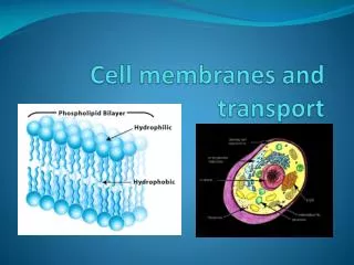
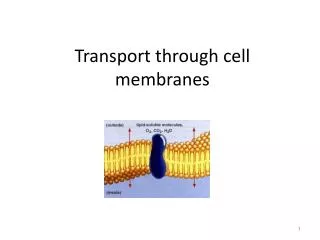
Phospholipids can form: BILAYERS -2 layers of phospholipids with hydrophobic tails protected inside by the hydrophilic heads. The PHOSPHOLIPID BILAYER is the basic structure of membranes. A Phospholipid Bilayer
Phospholipids Cell membranes are made mainly of phospholipids. They have: HYDROPHILIC heads (water liking) -Attracted to the water POLAR HYDROPHOBIC tails (water fearing) -Not attracted to the water NON-POLAR Phospholipids can form BILAYERS -2 layers of phospholipids with hydrophobic tails protected inside by the hydrophilic heads. The PHOSPHOLIPID BILAYER is the basic structure of membranes. Structure of the cell membrane
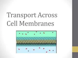
Diagram representing the cell membraneRemember the membrane is 7nm wide
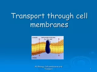
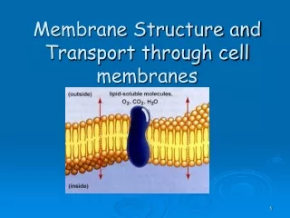
Fluid mosaic model Cell membranes also contain proteins within the phospholipid bilayer. This ‘model’ for the structure of the membrane is called the: FLUID MOSAIC MODEL FLUID- because individual phospholipids and proteins can move around freely within the layer, like it’s a liquid. MOSAIC- because of the pattern produced by the scattered protein molecules when the membrane is viewed from above.
The fracture occurs between the two phospholipid layers. You can clearly see the exposed proteins sticking out of the two layers. Individual phospholipids are too small to see. TEM of freeze-fractured cell membrane.
Cell Membranes from Opposing Neurons (TEM x436,740). } Phospholipid Bilayer 7nm wide
Features of the fluid mosaic model • Double layer – BILAYER of phospholipids which can move about by ………………………… in their own ………………………. • Phospholipid tails point inwards forming a ……. ………. ………………………… interior. The phospholipid heads point outwards facing the aqueous (water containing) medium surrounding the membrane. • Some phospholipids fatty acid tails are ……………………….. – straight so fit together tightly. Some are ………………………… – bent so fit together ……………………. The more unsaturated tails there are the more ……………… the membrane becomes. The lower the temp, the ……………….fluid. • Most protein molecules …………………. like icebergs in the layers, some are fixed to ………………………. inside the cell and don’t float. • Some proteins are embedded in the outer layer, some in the inner layer and some ………………… the two layers. Hydrophobic and Hyrdophilic parts of the protein molecules sit next to the …………………………… and ………………………….. portions of the ……………………………… of the membrane. This ensures the proteins stay in the membrane. • The membrane is ……nm thick on average. • Some phospholipids have carbohydrates attached to them called- ……………………….. • Some of the proteins have carbohydrates attached to them called– …………………… • The membrane also contains molecules of ………………………………..
Features of the fluid mosaic model • Double layer – BILAYER of phospholipids which can move about by DIFFUSION in their own MONOLAYER • Phospholipid tails point inwards forming a NON-POLAR HYDROPHOBIC interior. The phospholipid heads point outwards facing the aqueous (water containing) medium surrounding the membrane. • Some phospholipids fatty acid tails are SATURATED – straight so fit together tightly. Some are UNSATURATED – bent so fit together loosely. The more unsaturated tails there are the more ‘fluid’ the membrane becomes. The lower the temp, the less fluid. • Most protein molecules float like icebergs in the layers, some are fixed to structures inside the cell and don’t float. • Some proteins are embedded in the outer layer, some in the inner layer and some span the two layers. Hydrophobic and Hyrdophilic parts of the protein molecules sit next to the Hydrophobic and Hydrophilic portions of the phospholids of the membrane. This ensures the proteins stay in the membrane. • The membrane is 7nm thick on average. • Some phospholipids have carbohydrates attached to them – GLYCOLIPIDS • Some of the proteins have carbohydrates attached to them – GLYCOPROTEINS • The membrane also contains molecules of CHOLESTEROL
Roles of components of cell membranes Using the following headings produce a table on A4 to summarise roles of t he different types of molecules found in the cell membrane. Try not to copy, pick out the relevant information and write it in note form You may want to use bullet points, different coloured pens etc basically whatever helps you to remember them. There will be a short test on this next lesson! Use pages 53-54. Component Function • Phospholipids • Cholesterol • Proteins • Glycolipids and Glycoproteins
Summary • Cell membranes have a basic structure composed of a PHOSPHOLIPID BILAYER. • Phospholipds have HYDROPHOBIC (non-polar) tails and HYDROPHILIC (polar) heads. • The best model of the cell membrane is called the FLUID MOSAIC MODEL • The average thickness of the membrane is 7nm. • The fatty acid tails of phospholipids can be SATURATED (straight) or UNSATURATED (bent) • Proteins can float or be fixed and also have hydrophobic and hydrophilic portions. • Some proteins and phospholipids have carbohydrates attached to them to form GLYCOPROTEINS AND GLYCOLIPIDS. • Phospholipids form the bilayer, act as barrier to most water soluble substances • Cholesterol regulates the fluidity of the membrane, gives mechanical stability and help to prevent ions from passing through the membrane. • Proteins act as transport proteins to act as channels for substances to move into or out of the cell. Some act as membrane enzymes and some have important roles in membranes of organelles. • Glycolipids and Glycoproteins help to stabilise membrane structure, some act as receptor molecules eg for hormones and neurotransmitters or as antigens for other cells to recognise them.