Download
1 / 27
270 likes | 440 Views
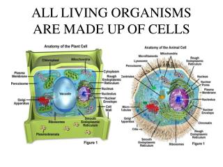
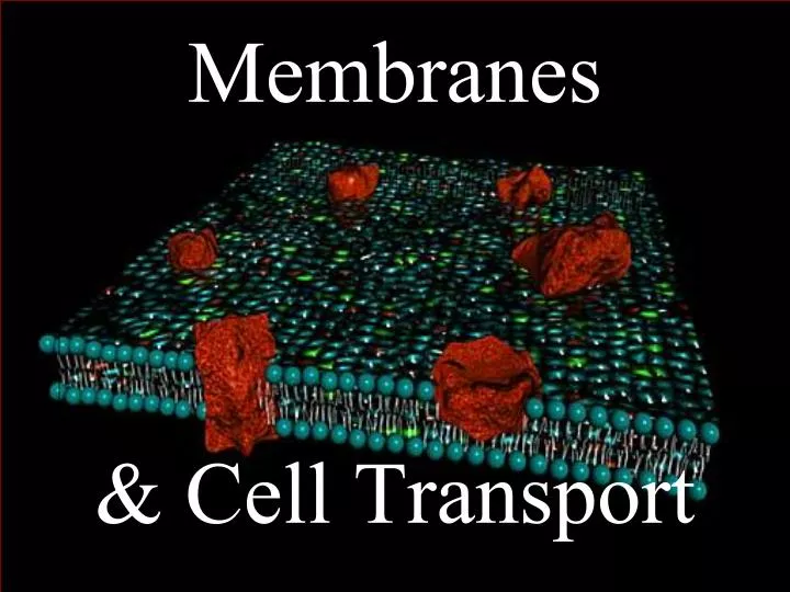
Membranes & Cell Transport. Cells lining intestinal tract. Blood cells. LE 3-1. Smooth muscle cell. Bone cell. Neuron in brain. Fat cell. Sperm. Ovum. Cilia. Secretory vesicles. Cytoplasm. LE 3-2. Mitochondrion. Plasma (cell) membrane. Nuclear envelope surrounding

E N D
Cells lining intestinal tract Blood cells LE 3-1 Smooth muscle cell Bone cell Neuron in brain Fat cell Sperm Ovum
Cilia Secretory vesicles Cytoplasm LE 3-2 Mitochondrion Plasma (cell) membrane Nuclear envelope surrounding nucleus Chromatin (DNA)
Form the boundary between the intracellularcompartment and the extracellular environment. What do membranes do? • “Traffic Cop” - Regulate what enters and leaves the cell = “selective permeability.” • Respond to substances that come in contact with the membrane. Ex: insulin, glucagon, & other hormones • Secrete (=squeeze out) substances that are synthesized inside the cell. • Compartmentalize and organize the interior of the cell. Ex: mitochondria, E.R., various vesicles
Early evidence for the bi-layered structure of the plasma membrane came from transmission electron micrographs. This is the plasma membrane of a RBC.
Here is a detailed picture of the way six phospholipid molecules interact with each other and their surroundings to form a phospholipid bilayer.
Phospholipid Animation (Click Here)
EXTRACELLULAR FLUID Proteins Phospholipid bilayer Protein with channel Carbohydrate chains Hydrophobic tails Cell membrane LE 3-3 Cholesterol Hydrophilic heads Proteins Cytoskeleton Protein with gated channel CYTOPLASM
EXTRACELLULAR FLUID Lipid-soluble molecules, O2 and CO2 diffuse through membrane lipids. Channel protein Plasma membrane LE 3-5 Small water-soluble molecules and ions diffuse through membrane channels Large molecules that cannot diffuse through lipids cannot cross the membrane unless they are transported by a carrier mechanism CYTOPLASM
Diffusion = spreading of molecules from a place where the concentration [ ] is higher to a place where it’s lower. LE 3-4
OSMOSIS = diffusion of H2O, across a membrane, from a region of higher [H2O] to a region of lower [H2O]. “[ ]” means “concentration of…”
Gray dots represent solute particles. Solute = anything dissolved in the water.
Two solutions containing different solute concentrations are separated by a selectively permeable membrane. Water molecules (small blue dots) begin to cross the membrane toward solution B, the solution with the higher concentration of solutes (larger pink circles). LE 3-6-1 A B Water molecules Glucose molecules Selectively permeable membrane
At equilibrium, the solute concentrations on the two sides of the membrane are equal. The volume of solution B has increased at the expense of that of solution A. LE 3-6-2a Volume increased Volume decreased
Diffusion & OsmosisAnimations http://www.biologycorner.com/bio1/diffusion.html http://www.tvdsb.on.ca/westmin/science/sbi3a1/Cells/Osmosis.htm http://www.stolaf.edu/people/giannini/flashanimat/transport/osmosis.swf
Water molecules LE 3-7a Isotonic
Water molecules LE 3-7b Hyp0tonic
Solute molecules LE 3-7c Hypertonic Hypertonic
Glucose molecule attaches to receptor site EXTRACELLULAR FLUID LE 3-8 Change in shape of carrier protein Receptor site Glucose released into cytoplasm Carrier protein CYTOPLASM
EXTRACELLULAR FLUID 3 Na+ LE 3-9 Sodium– potassium exchange pump 2 K+ ADP ATP CYTOPLASM
Ligands EXTRACELLULAR FLUID Ligands binding to receptors Endocytosis Exocytosis Ligand receptors LE 3-10 Coated vesicle CYTOPLASM Fusion Detachment Lysosome Fused vesicle and lysosome Ligands removed
Cell membrane of phagocytic cell Lysosomes LE 3-11 Vesicle Foreign object CYTOPLASM Pseudopodium (cytoplasmic extension) Undissolved residue EXTRACELLULAR FLUID
LE 3-12 Microvillus Microfilaments Cell membrane Mitochondrion Intermediate filaments Endoplasmic reticulum Secretory vesicle Microtubule
EXTRACELLULAR FLUID Endoplasmic reticulum CYTOSOL Lysosomes Cell membrane LE 3-14a Secretory vesicles Transport vesicle Golgi apparatus Membrane renewal vesicles Vesicle incorporation in cell membrane
LE 3-14b Exocytosis
http://www.wiley.com/legacy/college/boyer/0470003790/animations/membrane_transport/membrane_transport.htmhttp://www.wiley.com/legacy/college/boyer/0470003790/animations/membrane_transport/membrane_transport.htm Transport TypesAnimations