Download
1 / 19
190 likes | 218 Views
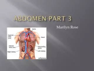
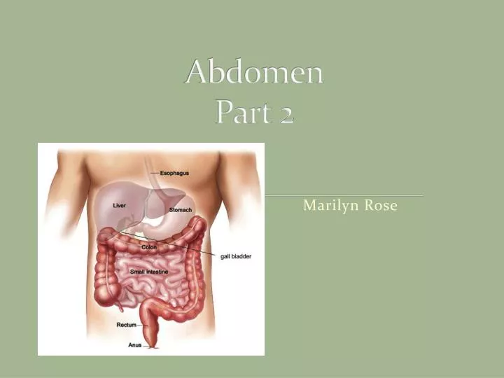
Abdomen Part 2. Marilyn Rose. Liver. Largest organ of abdomen Rt hypochondriac/ and epigastric regions Borders: Superior/lateral and anterior= Rt diaph Medial= sto/duodenum, transverse colon . Inferior= hepatic flex Posterior= Rt Kid Glisson’s Capsule Covered in peritoneum

E N D
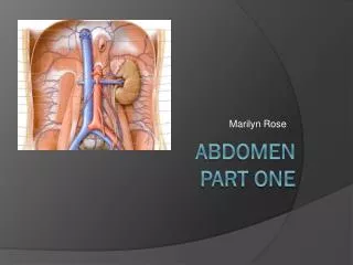
Abdomen Part 2 Marilyn Rose
Liver • Largest organ of abdomen • Rt hypochondriac/ and epigastric regions • Borders: • Superior/lateral and anterior= Rt diaph • Medial= sto/duodenum, transverse colon. • Inferior= hepatic flex • Posterior= Rt Kid • Glisson’s Capsule • Covered in peritoneum • Except: GB fossa, area of IVC and bare area.
Fissures of liver • Ligamentum teres- divides lt hepatic lobe into medial and lateral segments • Ligamentum venosum- separates the caudate lobe from Lt lobe • Transverse (portal) fissure- horizontal R/L PVS • Main lobar fissure (GB)- divides R/L lobes • Porta Hepatis- inferomedial border of the liver- the site of the MPV/HA and common bile duct.
Lobes of the liver • Lt lobe- • most anterior- lt of midline- separated from Rt by interlobar fissure/ Middle hepatic vein. • Caudate lobe • Smallest lobe, on the inferior/posterior surface, between the IVC and ligamentum venosum • Quadrate lobe- • Anterior-inferior surface of LT lobe between GB and ligamentum teres (remnant fetal umbilical vein, which runs along edge of falciform- The falciform supports/ attaches liver to diaphragm. 1: Right lobe of liver2: Left lobe of liver3: Quadrate lobe of liver4: Round ligament of liver5: Falciform ligament6: Caudate lobe of liver7: Inferior vena cava8: Common bile duct9: Hepatic artery10: Portal vein
Segments of Liver • Divided into 8 segments by the vascular supply (4=a/b) • Three hepatic veins: • MHV- divides liver into Rt/Lt lobes • RHV- divides Rt lobe into medial and lateral • LHV- Lt lobe into medial and lateral • Rt/ Lt Portal veins: • Divides each section transversely • Each segment is functionally independent with its own artery, portal vein and bile duct
Portal System • Nutrient rich blood from GI tract via the portal veins. • (75-80%) • Formed in the retroperitoneum by the superior mesenteric and splenic veins. • (posterior to the neck of the pancreas) • Porta hepatis- the portal vein branches into the Rt and Lt main portal veins- following the course of the HA’s • RPV branches into anterior and posterior branches • LPV courses left and then turns medially
End Stage Liver Failure Portal Hypertension-leads to Ascites and splenomegaly
Hepatic Artery • Arterial blood (20-25%) from common hepatic artery • Common hepatic artery arises from the celiac, entering the liver ANTERIOR to the portal vein. • The common hepatic artery arises from the celiac and branches into the Rt gastric and gastroduodenal arteries and continues ast eh proper heaptic towards the porta hepatis. • Prior to entering the liver it divides into the LT/RT hepatic arteries; which bring blood to each lobe.
Hepatic Veins IVC • Right Hepatic Vein • Largest- drains segments 5, 6 and 7 • Left Hepatic Vein • Smallest – drains segments 2,3 • Middle Hepatic Vein • Interlobar fissure- drains segments 4,5 and 8 • The three veins converge and enter the IVC just below the diaphragm
Biliary System • Gallbladder and bile ducts • Drain the liver and store bile until transported to the duodenum for digestion of fats.. • GB lives in the GB fossa- anterioinferior Rt lobe of the liver closest to the main lobar fissure. • Reservoir -fundus, body and neck- cystic duct Rt/Lt= CBD • Rt/Lt hepatic ducts unite at the porta hepatis to form the common hepatic duct (CHD), the CHD joins the cystic duct and forms the CBD. • CBD continues posterior to the pancreatic head and enters the duodenum along with the main pancreatic duct (Duct of Wirsung) at the Ampulla of Vater ( the muscle at the opening is called the sphincter of Oddi.
Biliary System Imaging ERCP MRCP Ultrasound
ERCP- Cholangiocarcinoma
Pancreas • Retroperitoneal, long, narrow- un-encapsulated • Posterior to the stomach/ between duodenum and splenic hilum • Head- located at the second portion of the duodenum about L2-L3, anterior to IVC and renal veins • Landmarks: • CBD Rt posterior and GDA on the anterior aspect • Uncinate process- between SMV and IVC • Neck- portal splenic confluence • main landmark- posterior to the neck • Body- largest, anterior to AO and SMA with the splenic vein running along the posterior surface • Tail- extends to the LT anterior pararenal space and LT kidney • Endocrine (insulin) and Exocrine (digestive enzymes) • Enzymes= amylase, lipase and peptidases and sodium bicarbonate
Spleen SAGITTAL • Largest lymph organ • Red pulp= blood • White pulp= lymphoid tissue and white blood cells • Intraperitoneal organ- covered in peritoneum • Posterior to stomach in the LUQ • Behind the 9-11th ribs • Borders- • medial- Lt kidney, splenic flexure, pancreatic tail • Posterior- diaphragm, pleura, Lt lung, ribs • Attached to > curvature of sto and Lt kid by the • Gastrosplenic and lienorenal ligaments TRANSVERSE
Spleen Splenic Rupture