Download
1 / 21
210 likes | 337 Views
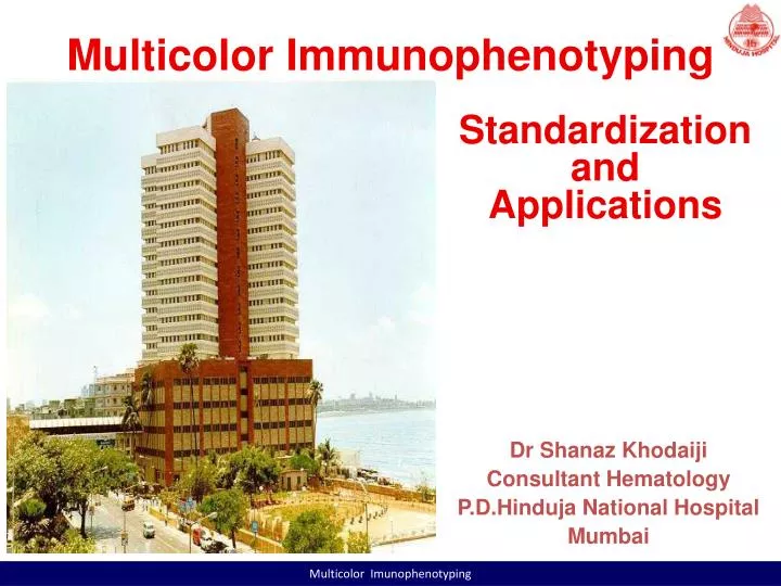
Multicolor Immunophenotyping. Standardization and Applications. Dr Shanaz Khodaiji Consultant Hematology P.D.Hinduja National Hospital Mumbai. Multicolor Imunophenotyping . History of FC at the PDHNH & MRC. FACSCalibur in 1995 FACSCanto II in 2008 CD4 and CD8 counts

E N D
Multicolor Immunophenotyping Standardization and Applications Dr Shanaz Khodaiji Consultant Hematology P.D.Hinduja National Hospital Mumbai Multicolor Imunophenotyping
History of FC at the PDHNH & MRC FACSCalibur in 1995 FACSCanto II in 2008 CD4 and CD8 counts Detailed Lymphocyte Subset analysis HLA-B27 Immunophenotyping of Acute Leukemias and CLPDs Platelet antigen studies Multicolor Imunophenotyping
Calibration using 7-colour setup beads • Prior to sample acquisition, the FC is set up using 7-Colour • setup beads. Controls need to address 3 issues Detectors, Thresholds, and Spectral Overlap. Beads are used • To adjust Detector Voltages: this ensures that fluorescence brightness is correct for stained cells in each detector. • Adjust the signal for events displayed in plots by changing detector voltages. Higher voltages amplify the signal. Lower voltages decrease the signal. • BD FACSCanto clinical software recalculates spectral overlap when you change detector voltages. Multicolor Imunophenotyping
BD FACS 7-colour setup beads Adjusting Thresholds • A threshold sets a channel number below which events will not be processed. Use threshold to filter out unwanted events. You can set one or more thresholds at a time, and choose whether any one or all need to be met. Adjusting Spectral Overlap • Set fluorescence compensation. Fluorochromes emit light over a range of wavelengths. During cytometer setup, fluorescence spillover is automatically determined and corrected. If necessary, you can use the spectral overlap controls to make manual adjustments. Multicolor Imunophenotyping
Monitor daily instrument performance • The LJ Chart for PMT Voltages and Blue and Red laser current are to be reviewed on a monthly basis to monitor Cytometer performance and see shifts or trends in parameters as they occur. The results are to be signed by the consultant and filed. • For the HLA B27 and Lymphocyte subset analysis the Lyse/No wash setup is used and for leukemia/lymphoma Immunophenotyping a lyse/wash setup is used. Multicolor Imunophenotyping
Cytometer setup report Multicolor Imunophenotyping
Optimization – why? Optimizing Cytometer Settings • When you performed cytometer QC, voltage settings were adjusted to set each parameter at a target value. These settings might not be appropriate for the stained samples you plan to analyze. Before recording data, adjust FSC, SSC, and threshold settings; gate on the population of interest (such as lymphocytes) and adjust voltages to optimize fluorescence signal. Multicolor Imunophenotyping
Reference range for Lymphocyte subsets Multicolor Imunophenotyping
Reference ranges in children Surg Cdr GauravNarula, Dr Shanaz Khodaiji, Col M.S. Bindra Multicolor Imunophenotyping
Comparative study of CD 4 counts (x 103/ml) Multicolor Imunophenotyping
Role of flow cytometric evaluation of lymphocyte subsets in predicting acute rejection episodes in renal transplant pts A. Dasgupta, S.Khodaiji et al • Aim of the study • To study short term results of renal transplant using low dose (1 ml/day) anti CD3 monoclonal antibody induction • to examine role of flow cytometricallydetermined lymphocyte counts in predicting ARE • Conclusion • Low dose Mab significantly decreases the CD3 count & CD4:CD8 ratio • FC allows monitoring of lympho subsets even at very low counts • AREs tend to be milder and later with use of Mab induction • CD3 counts< 10% and CD4:CD8 < 0.11 of baseline decrease the risk of ARE Multicolor Imunophenotyping
Flow cytometric analysis helps in the diagnosis of dense granule deficiency and qualitative deficiency of GPIIB/IIIA in platelets Sheeba Abraham & Amar Das Gupta • To correlate aggregometry & flow cytometry • findings • To evaluate the role of FC in diagnosis PFD Multicolor Imunophenotyping
Results and observations • Flow cytometry helped in distinguishing between quantitative • (5 cases) and qualitative (1 case) defects of platelet • GpIIb/IIIa in GT patients • In 4 patients with bleeding diathesis but normal • aggregation response, the diagnosis of SPD could be • made from flow cytometric demonstration of reduced • mepacrine uptake • Reduced P-selectin (CD62P) expression by flow cytometry • was seen as an isolated defect in 6 patients with near • normal aggregation response • FC analysis of platelet structure & function supplements • information obtained by aggregometry and helps in further • diagnosis & sub classification of platelet function disorders Multicolor Imunophenotyping
Flow cytometric estimation of CD41, CD61 for GT Multicolor Imunophenotyping
Comparison of platelet counts by Sysmex XE-2100 (I and O) and LH 750 with the International Flow Reference method in thrombocytopenic patients Multicolor Imunophenotyping
Analysis of Sysmex reported, Sysmex Impedence, Sysmex Optical and LH750 Methods with the IRM at Different Transfusion Thresholds Multicolor Imunophenotyping
PNH project Standardisation of Six colour Flowcytometry assay for the diagnosis and monitoring of Paroxysmal Nocturnal Hemoglobinuria using FLAER, as per 2010 International Guidelines RBCs- CD55/CD59/CD235a Multicolor Imunophenotyping
MRD for B ALL • MRD assay Standardisation based on the COG protocol • Normal samples/Non B ALL post induction Bone marrows /Staging marrows shall be used for making normal templates • Dilutional studies shall be done for linearity assessment and establishing cutoffs Multicolor Imunophenotyping
Why should we do it? FCM facilitates the analysis of cells within discrete subpopulations defined and selected (gated) based on other parameters, allowing a valid and reliable diagnosis, especially in NHLs and enabling their subclassification. FCM is fasterthan the histopathologic examination, allowing for therapeutic decisions to be made quickly. Allows a clear-cut correlation of multiple measurements (antigen expressions, DNA content, light scatter) in individual cells. Comparison of FC analysis on LNs with histopathologic study to diagnose NHL Multicolor Imunophenotyping
Thank You • Conducted workshops • and seminars • Training programs • Research projects • ILCP • CAP