Download
1 / 19
210 likes | 400 Views
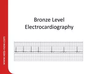
Bronze Level Electrocardiography. Aims. Brief summary of relevant clinical electrophysiology Indications for taking an electrocardiogram (ECG) How to obtain a diagnostic ECG Basic ECG interpretation. Section 1 – electrophysiology for clinicians. Unique properties of cardiomyocytes.

E N D
Aims • Brief summary of relevant clinical electrophysiology • Indications for taking an electrocardiogram (ECG) • How to obtain a diagnostic ECG • Basic ECG interpretation
Unique properties of cardiomyocytes • Electrical syncytium This means that the cells are coupled together in a way that permits rapid conduction of electrical impulses • Automaticity This describes the ability of cardiomyocytes to spontaneously depolarise. Under normal conditions the cells of the sinoatrial node have the fastest rate of spontaneous depolarisation and therefore are the dominant pacemaker cells.
What is the ECG measuring? • Electrical activity detected at the body surface • Cardiac tissue • Neuromuscular tissue (= movement) • Movement artefact such as trembling results in irregular baseline movement as shown below:
Einthoven’s triangle • Dr Einthoven invented the first practical ECG in 1903 • Einthoven’s triangle refers to the imaginary equilateral triangle formed by the 3 standard limb leads Left forelimb Left hindlimb - Lead III + - Lead II + + Lead I - The dots demonstrate the standard electrode positions
Anatomy of the intracardiac conduction system Bundle of His Sinoatrial node (SAN) Atrioventricular node (AVN) Left bundle branch Right bundle branch Right Left
Origin of -QRS-T P ECG: P -VE (Right forelimb in lead II) Wave of depolarisation moves from sinoatrial node across atria from right to left thereby creating a flow in current towards the positive electrode +VE (Left hindlimb in lead II) Right Left
Origin of P- RS-T Q ECG: P Q -VE (Right forelimb in lead II) Small delay as impulse traverses AVN hence trace returns to baseline. Depolarisation of the proximal interventricular septum then creates a small negative deflection – the Q wave. +VE (Left hindlimb in lead II) Right Left
Origin of P- Q S-T R ECG: P QR -VE (Right forelimb in lead II) Wave of depolarisation moves rapidly through the conduction system to the heart apex thereby creating a flow in current towards the positive electrode – the R wave +VE (Left hindlimb in lead II) Right Left
Origin of P- QR -T S ECG: P Q R S -VE (Right forelimb in lead II) Wave of depolarisation moves from the cardiac apex towards the heart base +VE (Left hindlimb in lead II) Right Left
Origin of P- QRS- T ECG: P QRS T -VE (Right forelimb in lead II) Wave of depolarisation moves from sinoatrial node across atria from right to left thereby creating a flow in current towards the positive electrode +VE (Left hindlimb in lead II) Right Left
Section 2 - Indications for obtaining an ECG • Common indications: • Document heart rate and rhythm • Dysrhythmia on auscultation • Less common indications: • Electrolyte abnormalities • Suspected drug toxicity • Suspected cardiac chamber enlargement
Patient set up for conscious ECG • Patient calm and still • Good electrical contact • Clips over bony areas to reduce muscle artefact • 50mm/s in leads I, II, III, aVL, aVR and aVF • 25mm/s rhythm strip for 1-5 minutes
Set up for monitoring ECG • Multi-parameter monitors • Tape ensures good contact between electrode and pad
Muscle movement artefact A common artefact seen on ECG is movement artefact caused by electrical activity present in moving muscles being detected by the ECG This results in rapidly undulating baseline movement which does not disrupt the superimposed heart rhythm.