Download
1 / 36
390 likes | 735 Views
Historical Perspective of EMG. Tiffany Zachry KIN 747 – Biomechanics Seminar Spring 2004. Or… Why Frogs Hate Scientists. Jan Swammerdam (1637-1680). Dutch anatomist and biologist Only one known portrait of him—and it’s a fake. Copied from Rembrandt’s The Anatomy Lesson of Dr. Tulp.

E N D
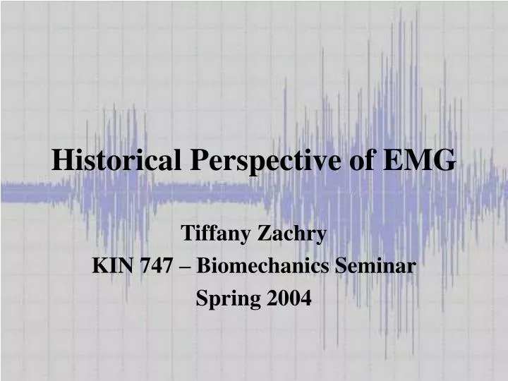
Historical Perspective of EMG Tiffany Zachry KIN 747 – Biomechanics Seminar Spring 2004
Jan Swammerdam (1637-1680) • Dutch anatomist and biologist • Only one known portrait of him—and it’s a fake. • Copied from Rembrandt’s The Anatomy Lesson of Dr. Tulp Picture Source: http://www.janswammerdam.net/
Discovered that stroking the innervating nerve of the frog’s m. gastrocnemius generated a contraction (1). Also strong evidence that he conducted the first electrical stimulation experiments, 134 years before Luigi Galvani (2). Swammerdam (cont’d)
Francesco Redi (1626-1698) • First to recognize connection between muscles and generation of electricity (1). • 1666—documented that electric ray fish used a highly-specialized muscle (3). • Most famous for establishing that maggots do not spontaneously generate from rotting meat. Picture Source: http://www.liberliber.it/biblioteca/r/redi/
Developed a device which produced electricity, which could be used to stimulate muscles. (3) Invented the first electric battery. The modern term “volt” comes from his name.Source: www.dictionary.com Alessandro Volta (1745-1827) Picture Source: http://www.th.physik.uni-frankfurt.de/~jr/physlist.html
Luigi Galvani • Credited as the father of neurophysiology for his similar work with frogs’ legs—1791 • Showed that “electrical stimulation of muscular tissue produces contraction and force.” (1) • Because of limited instrumentation, his work was not fully accepted until almost 40 years later. Picture Source: http://info.uibk.ac.at/c/c7/c704/museum/en/physicists/galvani.html Picture Source: http://butler.cc.tut.fi/~malmivuo/bem/bembook/01/01.htm
WARNING! The following picture is graphic in nature and may not be suitable for all audiences. Children, pregnant women, the elderly, and those with weak sphincters are strongly cautioned.
Modern Galvanized Frog Picture Source: http://www.soilmedia.org/artistprojects/hertz/
First practical galvanometer developed in early 1800s (3) Galvanometer – “An instrument used to detect, measure, and determine the direction of small electric currents by means of mechanical effects produced by a current-carrying coil in a magnetic field.” (Souce:http:/dictionary.reference.com/search?q=galvanometer) In 1838, Matteucci used one to show that bioelectricity is connected with muscular contraction (1) 1842 – demonstrated the existence of the action potential accompanying a frog’s muscle Carlo Matteucci Picture Source: http://perso.club-internet.fr/dspt/spirales.htm
1848 – first to detect electrical activity in voluntary muscle contractions of man (3) Had subjects place fingers in saline solution Removed skin to reduce transfer resistance (1) Detected signal through electrodes connected to galvanometer when subjects contracted muscles Emil Du Bois-Reymond (1818-1896) TAKE THAT!!!
1850 – applied electric stimulation to intact skeletal muscles (4). Interested in medical electricity for therapeutic purposes. (5) Systematically mapped out functions of nearly every facial muscle (3) Worked often with “the old man” who had little feeling in his face. (6) Guillaume Duchenne (1806-1875) Picture Source: http://chem.ch.huji.ac.il/~eugeniik/history/duchenne.html
WARNING! The following picture is graphic in nature and may not be suitable for all audiences. Children, pregnant women, the elderly, and those with weak sphincters are strongly cautioned.
Picture Source: http://chem.ch.huji.ac.il/~eugeniik/history/duchenne.html
Picture Source: http://chem.ch.huji.ac.il/~eugeniik/history/duchenne.html
Picture Source: http://chem.ch.huji.ac.il/~eugeniik/history/duchenne.html
Picture Source: http://chem.ch.huji.ac.il/~eugeniik/history/duchenne.html
Picture Source: http://chem.ch.huji.ac.il/~eugeniik/history/duchenne.html
Duchenne also discovered some rather surprising information.
The muscles around the eyes are only active during a genuine smile. • An insincere smile involves only the muscles of the mouth. (6) • So, everyone can tell when you’re faking it.
Knowledge of EMG developed as fast as technology could keep up. The term electromyography comes from Etienne Marey, who modified Lippman’s capillary electrometer (1876) as one of his many contributions to kinesiology. (1) It was used, much like his sphygmograph, to provide a graphic representation of a beating heart. (1) Other Notables
Forbes et al. were probably the first to use floating electrodes on a moving body. They used them to record EMG signals in elephants. (1) Forbes also used a CRT to amplify action potentials. (1) Willem Einthoven made a string galvanometer in 1903 and won the Nobel Prize for it. (1) It uses a thin conductor wire placed between two magnets. Other Notables
Modern Floating Electrode Picture Source: http://www.njit.edu/old/bme/Classes/Mr.Bergen/BME687/BME687%20-%20Electrodes.pdf
Adrian and Bronk developed the concentric needle electrode in 1929. (1) Used it primarily for researching motor control and muscle schemes. (1) Enabled detection in individual and small groups of muscle fibers. (4) Hypodermic needle with insulated wire in its barrel (4). Other Notables Picture Source: http://www.nihonkohden.com/products/supplies/emg-electrodes.html
Constructed the first electromyograph from 1942-44 at McGill University (Montreal Neurological Institute) (1). Also created a unipolar needle electrode. (4) Used his instruments to perform groundbreaking work with epilepsy and neurology and is a member of the Canadian Medical Hall of Fame(http://www.cdnmedhall.org) Herbert Jasper (1906-1999)
1962 – Basmajian compiles all of the known information about EMG. (4) Also created fine-wire electrodes (7) that were more comfortable than needles and could be used longer. (8) The book Muscles Alive becomes an invaluable tool in the field and is updated through five editions, the last Carlo De Luca. Founded International Society of Electrophysiological Kinesiology, ISEK, in 1965. (9) ISEK worked to create standards for EMG usage and reporting. John Basmajian Picture Source: http://www.vulvodynia.com/about.htm
Probably the most influential person in recent EMG history. Wrote the oft-cited paper “The Use of Surface Electromyography in Biomechanics.” (10) Cautioned against failing to understand EMG’s limitations. (10) Carlo J. De Luca Picture Source: http://nmrc.bu.edu/fac_staff/director/
“Electromyography is a seductive muse because it provides easy access to physiological processes that cause the muscle to generate force, produce movement, and accomplish the countless functions that allow us to interact with the world around us…To its detriment, electromyography is too easy to use and consequently too easy to abuse.” (10)
Our EMG System • In the Biomechanics Lab we are currently using a MyoSystem 2000 by Noraxon. • “Our internationally known patent-protected technology incorporates a signal processing technique that overcomes interference known as artifact in a signal. The result is a scientifically reliable surface assessment of dynamic muscle activity. This patent-protected signal processing technology is contained in all of our surface electromyography (SEMG) instrumentation and is recognized as the standard of excellence worldwide.” (Source: www.noraxon.com)
MyoSystem 2000 • Older version of the new 1400A. • Tethered system using bipolar electrodes. • New version offers USB 2.0 compatibility, thinner lightweight cables, and selectable bandwidths for surface or fine-wire electrode use. Source: www.noraxon.com
Highly conductive wet gel Superior adhesion Comfortable foam backing Unique offset concept High quality Ag/AgCl sensor Oblong shape for easy placement (juvenile) Blue Sensor Electrodes Adult Juvenile Source: www.ambuUSA.com
Other Manufacturers of EMG • NeuroDyne Medical Corporation • The Prometheus Group • Electronic Engineering Corporation • Motion Lab Systems, Inc. – makes equipment and software, including a package that is compatible with Vicon Clinical Manager and enables Vicon to display raw EMG data. (http://www.emgsrus.com) • There are numerous other manufacturers as well.
Current Studies Using EMG • “The effect of internal versus external focus of attention on EMG activity during basketball free-throws” • “Effects of focus of attention on take-off and landing strategies” • Both studies are using EMG to help assess the effects of attention focus on muscle activity. • Also, David Groh’s thesis will compare EMG activity in throwing versus Thera-Band use.
References • 1. Medved, V. (2001). Measurement of human locomotion. Boca Raton, FL: CRC Press. • 2. Clarys, J. P. (1994). Electrology and localized electrization revisited. Journal of Electromyography and Kinesiology, 4, 5-14. • 3. Cram, J. R., and Durie, M. D. (In press). The history of muscle dysfunction and SEMG. Journal of Applied Psychophysiology and Biofeedback. Retrieved February 28, 2004 from www.semg.org. • 4. Basmajian, J. V. (1978). Muscles alive: Their functions revealed by electromyography. 4th ed Baltimore: Williams and Wilkins. • 5. Licht, S. (1971). History of electrodiagnosis. In S. Licht (ed.), Electrodiagnosis and electromyography. New Haven, CT: Elizabeth Licht, Publisher. • Katz, E. Retrieved March 9, 2004 from http://chem.ch.huji.ac.il/~eugeniik/history/duchenne.html. • 7. Basmajian, J. V., and Stecko, G. (1962). A new bipolar electrode for electromyography: Journal of Applied Physiology. 17, 849. • 8. Whittle, M. W. (1999). Gait analysis: An introduction. 2nd ed. Oxford: Butterworth-Heinemann. • 9. The history of ISEK. Retrieved March 9, 2004 from http://isek.bu.edu. • 10. De Luca, C. J. (1997). The use of surface electromyography in biomechanics. Journal of Applied Biomechanics, 13, 135-163.
Other Resources Aminoff, M. J. (1978). Electromyography in clinical practice. Menlo Park, CA: Addison-Wesley Publishing. Dainty, D. A., and Norman, R. W. (eds.) (1987). Standardizing biomechanical testing in sport. Champaign, IL: Human Kinetics Publishers. Enoka, R. M. (1988). Neuromechanical basis of kinesiology. (2nd ed.) Champaign, IL: Human Kinetics. Kleissen, R. F. M., Buurke, J. H., Harlaar, J., and Zilvold, G. (1998). Electromyography in the biomechanical analysis of human movement and its clinical application. Gait and Posture, 8, 143-158. Latash, M. L. (1993). Control of human movement. Champaign, IL: Human Kinetics. Latash, M. L., and Zatsiorsky, V. M. (eds.) (2001). Classics in movement science. Champaign, IL: Human Kinetics. Loeb, G. E., and Gans, C. (1986). Electromyography for Experimentalists. Chicago: University of Chicago Press.