Download
1 / 17
230 likes | 469 Views
Organization of the Nervous System. Anatomy & Physiology Mrs. Halkuff. The nervous system is the master controlling and communicating system of the body. The nervous system has 3 main functions: Uses sensory receptors to monitor changes inside and outside of the body. ( Sensory Neurons )

E N D
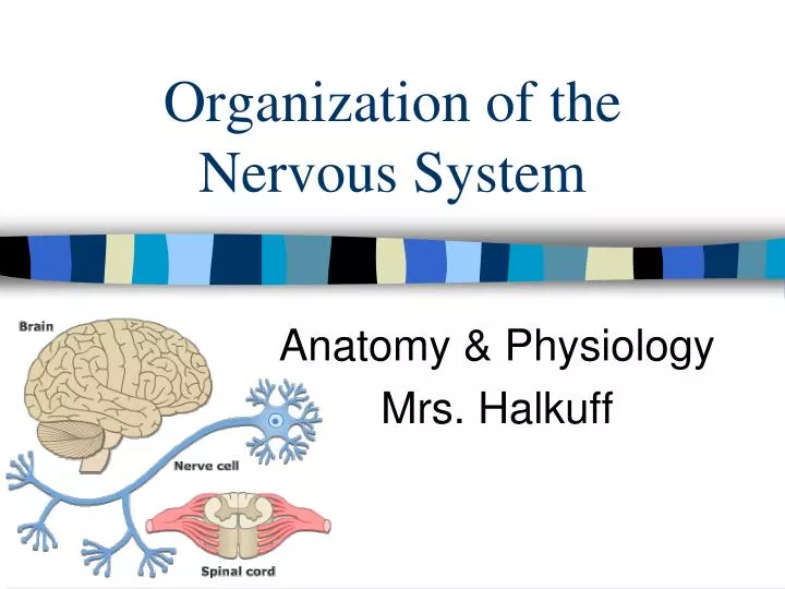
Organization of the Nervous System Anatomy & Physiology Mrs. Halkuff
The nervous system is the master controlling and communicating system of the body. • The nervous system has 3 main functions: • Uses sensory receptors to monitor changes inside and outside of the body. (Sensory Neurons) • Intergration: Processes and interprets sensory input and makes decision. • Motor output: Responds by muscles or glands. (Motor Neurons)
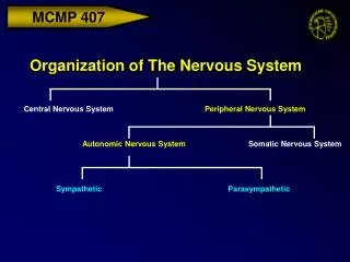
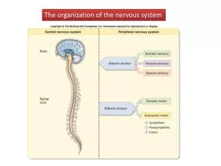
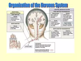
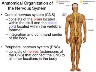
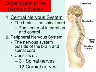
Organization of the Nervous System 1. Central Nervous System (CNS): • Brain and spinal cord • Command center • Interprets incoming sensory information • Make decisions based on past experiences
Organization of the Nervous System 2. Peripheral Nervous System (PNS): • Nerves that extend from the brain and spinal cord. 1. Sensory (Afferent) Division: Deliver impulses to the CNS from various parts of the body. 2. Motor (Efferent) Division: Carries impulses from the CNS to muscles and glands.
Neuron • Dendrites: Increase the surface area for receiving incoming information. • Axon: Carries information from the cell body to a neighboring neuron. • Myelin Sheath: Insulating fat cells that increase the rate of signal transmissions. • Node of Ranvier: Bare axon; allows action potential to jump from node to node. • Axon Terminals: Release chemicals called neurotransmitters.
Supporting Cells: CNS • 6 Cell Types Total: 4 CNS; 2 PNS • Microglia: Destroy invading microorganisms that could be harmful to the CNS. A type of macrophage. • Astrocytes: Most abundant; Anchors the neurons in place by attaching to capillaries. Also serve as a nutrient (blood supply) to neurons. • Ependymal Cell: Line the brain & spinal cord cavities (dorsal). Have cilia that help to circulate the cerebro-spinal fluid. • Oligodendrocytes: Wrap around axons of neurons to form myelin sheaths.
Supporting Cells: PNS • Schwann Cells: Help form myelin sheath; also engulf deteriorating cell debris & aid in regeneration. • Satellite Cells: Surround the cell bodies and regulate chemical environment.
Resting Potential • A neuron sends messages electrochemically. • Ions are Na & K (positive) • A neuron is at rest when it is not sending a signal and is in a negatively charged state. • Even at rest, the neuron allows K to pass. • Neuron pumps 3 Na ions out for every 2 K ions it pumps in. • At rest, there are more Na ions outside and more K ions inside • Resting & Action Potential
Action Potential • Occurs when a neuron sends information down the axon. • Electrical activity created by a depolarizing current. • A stimulus must make the neuron reach its threshold in order to fire an action potential. • Stimulus causes Na channels to open and Na+ rushes into the neuron, depolarizing it. • K rushes out of the cell, reversing the depolarization.
Autonomic Nervous System • Part of the PNS. • Has 2 divisions: Sympathetic & Parasympathetic • Controls heart rate, digestion, respiration rate, salivation, & perspiration.
Sympathetic • Neurons begin in the Thoracic & Lumbar region of the spinal cord • Functions in actions that require a quick response. • “Fight or Flight” response.
Parasympathetic • Neurons begin in the cervical & sacral regions of the spinal cord. • Functions in actions that do not require an immediate response. • “Rest & Digest” • Constant opposition to Sympathetic N.S. • Sympathetic & Parasympathetic Clip
Reflexes • Involuntary, rapid actions; usually for survival. • Most reflexes don’t have to travel to the brain, as they need to happen quickly. • Reflex Arc: • Receptors are excited. • Signal travels along sensory neuron to spinal cord • Signal is passed onto a motor neuron • Muscle/Gland is stimulated.