Download
1 / 19
210 likes | 622 Views
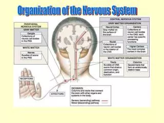
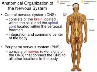
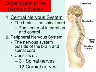
Organization of the Nervous System. Major Divisions of the Nervous System. External environment. Internal environment. Sensory input. Motor output. A “system of twos”. CNS: Bones and Meninges. Meninges. www.studyblue.com.

E N D
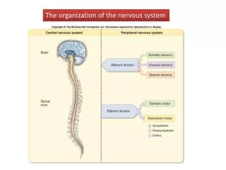
Organization of the Nervous System
Major Divisions of the Nervous System External environment Internal environment Sensory input Motor output A “system of twos”
CNS: Bones and Meninges Meninges www.studyblue.com The brain and spinal cord are the most protected organs in the body. They are encased in bone (skull and vertebrae) and covered by three meninges or membranes: dura mater; arachnoid mater; and pia mater.
CNS: Cerebrospinal Fluid Cerebrospinal fluid (CSF), fills the sub-arachnoid space covering the brain and spinal cord, the cerebral ventricles (four internal chambers of the brain) and the central canal of the spinal cord. CSF supports the CNS and provides cushioning against injury. en.wikipedia.org
CNS: Blood Flow 1 en.wikipedia.org The brain is finely tuned electrochemical organ with substantial nutritional requirements. Blood is supplied from three main arteries: the anterior, middle and posterior cerebral arteries
CNS: Blood Flow 2 The spinal cord is supplied by three main arteries that run longitudinally (top to bottom): the anterior and the right and left posterior arteries. www.studyblue.com
CNS: Blood Barriers Blood-brain barrier (BBB) Blood-spinal cord barrier (BSCB) The CNS requires a constant environment to function properly Barriers, in the form of tightly packed endothelial cells lining blood vessel walls, maintain this environment by impeding passage into the CNS of: • "foreign substances" • proteins/other large molecules • highly charged molecules • hormones and neurotransmitters Glucose is actively transported The barrier is weak in some areas in the brain to allow monitoring of the chemical composition of blood
Directions in the Vertebrate Nervous System SUPERIOR CAUDAL ROSTRAL PROXIMAL DISTAL INFERIOR Directions in the vertebrate nervous system are described in relation to the orientation of the spinal cord in the standard anatomical position.
Anatomical Directions in the Human In humans, the directions in the cerebral hemispheres are rotated by 90°in comparison to those in the spinal cord and brain stem because of the unusual upright posture of humans. Thus, for example, the top of the head and the back of the body are both dorsal even though the directions are different.
Planes of Section Sections of the brain are usually shown in one of three orientations: • horizontal; • frontal (coronal); • sagittal (a midsagittal cut separates the left and right halves of the brain) Cross-section: cut at a right angle to a long narrow structure (e.g., spinal cord)
Five Major Divisions of the Brain Brainstem (Medulla) Divisions are based on developmental origins; they do not sub-serve discrete functions.
Spinal Cord 1 The spinal cord is located in the vertebral canal and is made up of 31 segments: 8 cervical; 12 thoracic; 5 lumbar; 5 sacral;1 coccygeal A pair of spinal nerves leaves each segment. The cord is shorter than the bony spinal column. The lower nerves run down the canal before exiting (cauda equina, “horse tail”) www.studyblue.com
Spinal Cord 2 DORSAL In cross section, it is apparent that the spinal cord comprises two different areas: • inner H-shaped core of gray matter (cell bodies) • outer area of white matter (myelinated axons) cnx.org VENTRAL From anterior to posterior: • white matter decreases • gray matter shows two enlargements (C5, L4) in the ventral horn for arms and legs www.washington.edu
Spinal Cord 3 Pairs of spinal nerves are attached to the spinal cord – one on the left and one on the right – at 31 levels of the cord: • dorsal, afferent, sensory • ventral, efferent, motor
Spinal Cord 4 Spinal cord grey matter contains a number of prominent nuclear groups. The white matter is organized into different ascending and descending tracts. thebrain.mcgill.ca www.wcc.hawaii.edu
Peripheral Nervous System Autonomic Somatic The branch of the NS of which we are conscious. It provides sensory and motor innervation to all body parts except organs, smooth muscles and glands. It is involved in sensations that we are aware of such as light and pain, and our voluntary movements. The branch of the NS of which we are unconscious. It regulates the visceral (organ) functions that maintain homeostasis within the body, including heart rate, blood pressure, digestion, etc. It has two efferent components in balance: sympathetic and parasympathetic.
Somatic Nervous System www.studyblue.com Somatic spinal nerves innervate a particular region of skin.
Autonomic Nervous System www.humankinetics.com Second stage neurons are near the target organ Second-stage neurons are far from the target
The Cranial Nerves “On Occasion Our Trusty Truck Acts Funny, Very Good Vehicle Any How” “Some Say Marry Money, But My Brother Says Big Brains Matter Most”