Download
1 / 27
270 likes | 974 Views
Case Presentation. XX is a 44 yo WF referred to hematology for evaluation of hypoxemia and possible carbon monoxide poisoningShe was admitted with a 5 day history of dyspnea, productive cough, chest pain and fever, at which time she had a Pa O2 of 74 on ABG and was cyanoticAlthough Pa O2 increas

E N D
1. A patient with hypoxemia and cyanosis Kathy Ponder
2. Case Presentation XX is a 44 yo WF referred to hematology for evaluation of hypoxemia and possible carbon monoxide poisoning
She was admitted with a 5 day history of dyspnea, productive cough, chest pain and fever, at which time she had a Pa O2 of 74 on ABG and was cyanotic
Although Pa O2 increased to 189 with oxygen, she remained cyanotic in the ICU with a high carboxyHb and hematology was consulted
3. Past Medical History Diagnosis of polycythemia vera a few years ago being treated with phlebotomy
History of carbon monoxide poisoning in 9-00 when she presented with dyspnea and was found to have high carboxy Hb; she was treated with hyperbaric oxygen; attributed to being a truck driver
Hepatitis C being treated with interferon and ribavarin
4. Medications Lorazepam
Paxil for depression
Interferon for Hepatitis C
Ribavirin for Hepatitis C
5. Social History Generally smokes 2 to 3 ppd, but had decreased to 1 ppd
Worked previously as truck driver but has not worked while on interferon
6. Family History No children
The patient has 3 siblings all of whom are normal
Mom has been blue all her life
Mom had 5 siblings, 4 of whom are blue
The patient had several maternal cousins that are blue; many were receiving phlebotomy for polycythemia vera
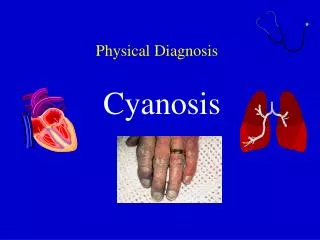
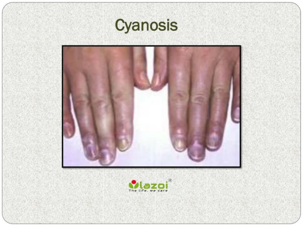
7. Physical exam WDWN female in mild respiratory distress
T=37.4, RR 24, P 88
Chest: Decreased BS at bases, R>L, occasional wheeze
The patient was cyanotic in the mucosa and extremites
8. Laboratories CBC: WBC 4.2, Hb 16.1 Hct 46.8, Platelets 267,000; MCV 101.9, RDW 14.9
Retic count 7.2% (0.5 to 1.5%)
LDH 204 (100 to 250)
9. ABGs Initial value on room air
7.42/34/74/23
Oxy Hb 71.5%
Met Hb 1.1%
Carboxy Hb 25%
Pulse oximetry at this time had 88% O2 sat Values on 100% oxygen
7.37/38/189/23
Oxy Hb 81.4%
Met Hb 1.4%
Carboxy Hb 16.5%
10. Additional Labs Drug screen: Negative
CXR: Diffuse fine reticular infiltrates
Chest CT: Negative for PE; multiple round soft tissue densities in RUL, RLL, LLL c/w infection or malignancy
11. What tests should be ordered, what does she probably have, and how should she be treated?
12. Hemoglobin Analysis Hemoglobin electrophoresis at Mayo
89% hemoglobin A (95%-98%)
2.6% hemoglobin A2 (2%-3.3%)
0.9% hemoglobin F (0%-2%)
7.3% Hb M-Saskatoon
Methemoglobin evaluation at Mayo
1.8% methemoglobin (0-1.5%); may decline 40% per day
1.3% sufhemoglobin (0-1%)
13. Labs, continued Hemoglobin reductase B:
8.5 IU/g Hb (normal 10.1 to 19.4)
ABG at Childrens (continuous CO-oximeter (ABL 735)
Oxy Hb 74%
Carboxy Hb 3.1%
Met Hb 5.0%
There were flags everywhere as the absorbance spectrum was unusual
14. Etiology of Met Hemoglobin Methemoglobin; autosomal dominant
Cytochrome b5 reductase deficiency; autosomal recessive
Drug ingestion
15. Hemoglobin MSaskatoon Histidine to tyrosine missense mutation at position 63 of b chain (Gerald and Efron; PNAS 47:1758, 1961)
This has increased oxygen affinity
Cyanosis due to methemoglobin
Can see mild hemolytic anemia, which can be exacerbated by sulfonamides
16. Met Hemoglobins Hb MBoston a58(E7) His to Tyr
Hb MSaskatoon b63 (E7) His to Tyr
Hb MIwate a87(F8) His to Tyr
Hb MHyde Park b92(F8) His to Tyr
Hb MMilwaukee b67(E11) Val to Glu
Hb FMFort Ripley g92(F8) His to Tyr
Hb FMOsaka g63 (E7) His to Tyr
18. Putative Chemistry of Tyrosine (in Met Hb) Fe+3 Interaction
19. Biochemistry of Met Hemoglobin Occasionally oxygen leaves the hemoglobin as a superoxide anion removing an electron from the Fe to oxidized it to Fe+3, producing methemoglobin, which cannot bind to molecular oxygen
Fe+3 is usually reduced to Fe+2 by the cellular machinery (cytochrome b5 reductase and cytochrome b5)
The His to Tyr mutations result in a tyrosine near the Fe, which forms an iron-phenolate complex that resists reduction by the cellular machinery, resulting in increased levels of methemoglobin
This results in a marked shift of the Hb dissociation curve and low pO2, since it is not possible to fully oxygenate the hemoglobin M
Cyanosis is due to the altered absorbance spectrum of methemoglobvin compared with oxyhemoglobin
20. Pulse Oximetry is not Accurate in Met Hb (Shannon Haymond, Rohit Cariappa, Chuck Eby, Mitchell Scott) Pulse oximetry measures absorbance at 660 nm and 940 nm
DeoxyHb has 3-fold more absorbance at 660 nm than at 940 nm
OxyHb has half as much absorbance at 660 nm as at 940 nm
Ratio of the red (660 nm) to near-infrared (940 is used to determine the % saturation
Met Hb has equal absorbance at 660 nm and 940 nm
Pulse oximetry is not reliable in the presence of dyshemoglobins; it can overestimate the oxygen saturation
21. Blood Gas Analysis in BJC lab for Hb MSaskatoon The absorption of Hb MSaskatoon differs from that of Met Hb (peaks at 500 and 631), resulting in a low Met Hb on blood gas analysis
The absorption of Hb M (peak at 600 nm) overlaps with that of carboxyHb, accounting for the high CO Hb on this admission and the prior admission when she received hyperbaric oxygen
22. Clinical Symptoms of HbM Present with cyanosis
Generally asymptomatic
Sometimes mild hemolysis
23. Deficiency of Cytochrome B5 Reductase or Cytochrome B5 Autosomal recessive
Results in over half of the cases of methemoglobin
Results in inability to maintain hemoglobin in reduced form
85% to 90% due to deficiency in RBC only (type I)
15% to 10% involve deficiency throughout the body (Type II, involve neurological symptoms)
Type IV is due to deficiency in cytochrome 5
Heterozygotes are asymptomatic but have an increased chance of symptoms with toxin exposure
Usually modest levels of Met Hb (10% to 20%) without symptoms
24. Ingestion of drugs that cause Methemoglobin Drugs
Dapsone
Primaquine
Nitrate
Phenacetin
Benzocaine
Amyl nitrite
Nitroglycerin
Nitroprusside
Chloroquinone
Sulfa
Chemicals
Nitrites
Hydrazine
Hydrazides
Thiols
Phenylenediamine
Aminochenols
Chlorobenzene
Aniline dye (in shoe dyes)
25. Correlation of Met Hb and Symptoms Met Hb 15% to 20%: Asymptomatic cyanosis
Met Hb >20%: Symptoms of dyspnea, fatigue, nausea, dizziness, HA, and syncope
Met Hb >70%: High mortality
26. Therapy Usually not indicated for Hb M or cytochrome B5 reductase deficiency
Can give methylene blue 1 mg/kg IV for drug ingestion if >20% Met Hb and the patient is symptomatic, or >30% Met Hb
Patients that are G6PD deficient (which reduces NADP+) do not respond well to methylene blue
Ascorbic acid at 500 to 1000 mg/day po
Riboflavin 20 to 60 mg/ day po
27. Plan No treatment for carbon monoxide poisoning
Discontinue phlebotomy as the polycythemia was due to low oxygen delivery
Home O2 (although not clear she needs it; repeat ABG PaO2 was 86% and 89% on room air)
Avoid sulfa
Stop smoking