Download

1 / 38
390 likes | 1.16k Views
A Patient with Pancytopenia. HISTORY: Part 1. A 34 year-old woman is seen by her dentist for bleeding from her gums. The bleeding started on the day prior to evaluation and seems to be getting steadily worse with time.

E N D
HISTORY: Part 1 • A 34 year-old woman is seen by her dentist for bleeding from her gums. The bleeding started on the day prior to evaluation and seems to be getting steadily worse with time. • After briefly evaluating the patient and noting diffuse bleeding from the mucosal surfaces around multiple teeth, the dentist refers her for further evaluation to the emergency department at a nearby hospital where you are working.
What further information should you request regarding the patient's history? 可以從下一頁來做選擇
Have you ever had a similar episode in the past? • Is there any family history of a bleeding disorder? • Are you taking any prescribed or over-the-counter medications? • Do you smoke cigarettes or drink alcohol? • Do you have any bruises or other areas of bleeding? • Is anyone else in your family ill? • Do you have any pets?
Answer • Have you ever had a similar episode in the past? • Is there any family history of a bleeding disorder? • Are you taking any prescribed or over-the-counter medications? • Do you smoke cigarettes or drink alcohol? • Do you have any bruises or other areas of bleeding? • Is anyone else in your family ill? • Do you have any pets?
HISTORY: Part 2 • The patient notes that she has never had an episode of anything similar in the past. Her past medical history is remarkable only for a tonsillectomy at age 11 that was entirely uncomplicated. There is no family history of a bleeding disorder. She takes no medication regularly, but on occasion takes acetaminophen for headaches. She does not smoke or drink alcohol.
HISTORY: Part 2 • On a review of systems, you discover that over the past month she has been feeling gradually more fatigued, which she attributes to working overtime at her job as an administrative assistant. She notes no fevers or chills, but does say that she may have lost a few pounds unintentionally due to decreased appetite. She notes that on her way to the emergency department, she noted some small bruises on her forearms and thighs and that her ankles and feet seem to have numerous small red dots on them. She denies any chest pain, shortness of breath, abdominal pain, edema, headaches, dizziness, or other neurologic symptoms. • After obtaining this information, you move on to perform a physical examination on the patient.
Any Question? Discussion
Discussion Can you list differential diagnosis according to the history and PE?
What additional information would you request at this time? Laboratory Data Radiologic Studies
Answer • Laboratory Data • Radiologic Studies
What laboratory data should you request initially? • CBC with differential • Peripheral Blood Smear • Prothrombin Time (PT) • Activated Partial Thromboplastin Time (aPTT) • Serum creatinine • Liver function tests
Answer • CBC with differential • Peripheral Blood Smear • Prothrombin Time (PT) • Activated Partial Thromboplastin Time (aPTT) • Serum creatinine • Liver function tests
Discussion What is the finding of the lab data? What is your differential diagnosis?
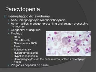
Peripheral blood smear: Review of the peripheral blood smear reveals decreased platelets, 3 to 4 schistocytes per high powered field, and a population of cells such as the one shown above.
III. LABORATORY DATA Are there further laboratory data, radiographic studies, or diagnostic procedures that you would wish to request at this time?
Fibrinogen level • LDH • Uric Acid • CT scan of the chest, abdomen, and pelvis • Lumbar puncture • Bone marrow aspirate and biopsy
Answer • Fibrinogen level • LDH • Uric Acid • CT scan of the chest, abdomen, and pelvis • Lumbar puncture • Bone marrow aspirate and biopsy
While the bone marrow aspirate and biopsy is being processed, what is the most likely diagnosis for this patient based upon the all of the data provided? • Aplastic anemia • Paroxysmal nocturnal hemoglobinuria • Sepsis syndrome • Vitamin B12 deficiency • Acute lymphoid leukemia • Acute myeloid leukemia
Discussion About your differential diagnosis
Answer • Aplastic anemia • Paroxysmal nocturnal hemoglobinuria • Sepsis syndrome • Vitamin B12 deficiency • Acute lymphoid leukemia • Acute myeloid leukemia
Acute myeloid leukemia may present with pancytopenia, leukocytosis, or a white blood cell count within the normal range. In addition, sometimes when pancytopenia is present, there are no circulating blasts, although many may be found in the bone marrow (this is called aleukemic leukemia). In this case, the combination of pancytopenia with blasts combined with the consumptive coagulopathy (disseminated intravascular coagulation, DIC) indicates a diagnosis of acute myeloid leukemia. The cell shown in the photomicrograph of the peripheral blood is a myeloblast that has a heavily granulated cytoplasm. The stick-like inclusions in the cytoplasm are Auer rods. These are stacks of lysosomes that have collapsed on one another to form linear structures within the cytoplasm. They are indicative of a myeloid disorder and are seen only in acute myeloid leukemia and advanced myelodysplastic syndromes. The latter are disorders that tend to transform into acute myeloid leukemia.
The bone marrow aspirate and biopsy findings become available, along with the flow cytometric analysis documenting acute promyelocytic leukemia (APL, also known as M3 AML by the older French-American British Classification scheme):
Overall, the blasts present in bone marrow are found to have the following phenotype: Present : CD33 CD13 CD117 Absent : CD34, HLA-DR • The presence of CD33 and absence of CD34 and HLA-DR are consistent with a diagnosis of acute promyelocytic leukemia (APL). The particular markers present also distinguish acute lymphoid from acute myeloid leukemia and also distinguish different classes of myeloid leukemias from one another. • Cytogenetic Analysis including Fluorescence in situ Hybridization (FISH) • Cytogenetic analysis and FISH analysis have become standard techniques in the analysis of acute leukemia. Cytogenetic abnormalities, present in about 50% of cases of acute myeloid leukemia, can have major prognostic implications. Cytogenetic abnormalities also have a major impact on treatment decisions, including the type of chemotherapy administered and whether or not to recommend hematopoietic stem cell transplantation in certain cases.
Giemsa banding of 20 metaphase chromosome spreads documents the presence of t(15;17) translocation in 18 cells. FISH demonstrates the presence of a PML/RARA signal in 85% of 200 interphase nuclei examined. • Molecular Diagnostics • Using reverse transcriptase polymerase chain reaction (RT-PCR) on RNA prepared from the patient’s white blood cells, a strong band is identified, indicating the presence of the PML/RARA gene fusion. • Molecular diagnostic testing is becoming increasingly important in leukemia diagnosis. Besides facilitating rapid diagnosis of known translocations, it identifies abnormalities in about 75% of acute myeloid leukemia that is otherwise cytogenetically normal. As with cytogenetics, identification of such abnormalities can have major prognostic significance. For example, in otherwise cytogenetically normal acute myeloid leukemia, the finding of internal tandem duplications in the FMS-like tyrosine kinase 3 gene (FLT3-ITD mutation) is associated with a poor prognosis, whereas the finding of an isolated abnormality in the nucleophosmin 1 gene (NPM1 mutation) is associated with a favorable overall prognosis.
The molecular pathogenesis of APL is now understood and is illustrated below. The fusion protein consists of the promyelocytic leukemia gene (PML), a tumor suppressor normally found in nuclear bodies, and the retinoid acid receptor alpha gene (RARA), a transcription factor that is normally responsive to retinoid acid. The juxtaposition of these two genes produces a protein that does not respond to the usual form of retinoic. However, it does respond to a specific type of retinoic acid, all-trans retinoid acid, leading to restoration of gene transcription and thereby to cellular differentiation. Interestingly, the substance arsenic trioxide leads to a similar effect because it leads to aggregation and degradation of the PML-RARA fusion protein, thereby promoting differentiation. Both ATRA and arsenic trioxide are now used for the treatment of APL.
All-trans retinoid acid • Daunomycin • Maintenance chemotherapy • Management of DIC • Follow up BM with PML/RARa to detect minimal residual disease
Discussion About treatment
Reference: ASH teaching case • http://teachingcases.hematology.org/