Download
1 / 51
620 likes | 989 Views
REGENERATION. HEALING (repair). LEARNING OBJECTIVES. Review the normal physiology and concepts of cell proliferation , cell growth, cell “cycle”, and cell differentiation Understand the basic factors of tissue regeneration

E N D
REGENERATION HEALING (repair)
LEARNING OBJECTIVES • Review the normal physiology and concepts of cell proliferation, cell growth, cell “cycle”, and cell differentiation • Understand the basic factors of tissue regeneration • Understand the relationships between cells and their ExtraCellular Matrix (ECM) • Understand the roles of the major players of healing---angiogenesis, growth factors (GFs), and fibrosis • Differentiate 1st & 2nd intention healing
DEFINITIONS: • REGENERATION: Growth of cells to replace lost tissues • HEALING: A reparative tissue response to a wound, inflammation or necrosis, often leads to fibrosis • GRANULATION TISSUE • “ORGANIZING” INFLAMATION
REGENERATION • Replacement of lost structures • Is dependent on the type of normal turnover the original tissue has • Can be differentiated from “compensatory” growth
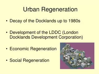
HEALING (repair) • Needs a wound, inflammatory process, or necrosis • Many disease appearances anatomically are the result of “healing” such as atherosclerosis • Often ends with a scar • Fibrosis, as one of the 3 possible outcomes of inflammation, follows “healing” • Requires a connective tissue “scaffold” • Fibrosis occurs in proportion to the damage of the ECM
Cell Population Fates • PROLIFERATION • Hormonal, especially steroid hormones • eg., EPO, CSF • DIFFERENTIATION* • UNIDIRECTIONAL, GAIN and LOSS • APOPTOSIS *One of the most KEY concepts in neoplasia
ECTODERM MESODERM ENTODERM
CELL CYCLE • G0 • Quiescent (not a very long or dominent phase) • G1 • PRE-synthetic, but cell GROWTH taking place • S • Cells which have continuous “turnover” have longer, or larger S-phases, i.e., DNA synthesis • S-phase of TUMOR CELLS can be prognostic • G2 • PRE-mitotic • M (Mitotic:, P,M,A,T, Cytokinesis)
CELL TYPES • Labile: eg., marrow, GI • Quiescent: liver, kidney • NON-mitotic: neuron, striated muscle
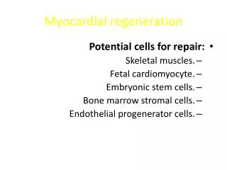
STEM CELLS(TOTIPOTENTIAL*) • EMBRYONIC • ADULT
EMBRYONICSTEM CELLS • DIFFERENTIATION • KNOCKOUT MICE (mice raised with specific gene defects) • REPOPULATION OF DAMAGED TISSUES, in research
ADULTSTEM CELLS • MARROW (HEMOCYTOBLAST) (hematopoetic stem cells) • NON-MARROW (RESERVE)
ADULT TISSUE DIFFERENTIATION and REGENERATION PARALLELS EMBRYONIC DEVELOPMENT
Growth Factors (GFs) • Polypeptides • Cytokines • LOCOMOTION • CONTRACTILITY • DIFFERENTIATION • ANGIOGENESIS
Growth Factors (GFs) • Epidermal • Transforming (alpha, beta) • Hepatocyte • Vascular Endothelial • Platelet Derived • Fibroblast • Keratinocyte • Cytokines (TNF, IL-1, Interferons)
CELL PLAYERS (source AND targets) • Lymphocytes, especially T-cells • Macrophages • Platelets • Endothelial cells • Fibroblasts • Keratinocytes • “Mesenchymal” cells • Smooth muscle cells
E (Epidermal) GF • Made in platelets, macrophages • Present in saliva, milk, urine, plasma • Acts on keratinocytes to migrate, divide • Acts on fibroblasts to produce “granulation” tissue
T (Transforming) GF-alpha • Made in macrophages, T-cells, keratinocytes • Similar to EGF, also effect on hepatocytes
H (Hepatocyte) GF • Made in “mesenchymal” cells • Proliferation of epithelium, endothelium, hepatocytes • Effect on cell “motility”
VE (Vascular Endothelial) GF • Made in mesenchymal cells • Triggered by HYPOXIA • Increases vascular permeability • Mitogenic for endothelial cells • KEY substance in promoting “granulation” tissue
PD (Platelet Derived) GF • Made in platelets, but also MANY other cell types • Chemotactic for MANY cells • Mitogen for fibroblasts • Angiogenesis • Another KEY player in granulation tissue
F (Fibroblast) GF • Made in MANY cells • Chemotactic and mitogenic, for fibroblasts and keratinocytes • Re-epithelialization • Angiogenesis, wound contraction • Hematopoesis • Cardiac/Skeletal (striated) muscle
T (Transforming) GF-beta • Made in MANY CELLS • Chemotactic for PMNs and MANY other types of cells • Inhibits epithelial cells • Fibrogenic • Anti-Inflammatory
K (Keratinocyte) GF • Made in fibroblasts • Stimulates keratinocytes: • Migration • Proliferation • Differentiation
I (Insulin-like) GF-1 • Made in macrophages, fibroblasts • Stimulates: • Sulfated proteoglycans • Collagen • Keratinocyte migration • Fibroblast proliferation • Action similar to GH (Pituitary Growth Hormone)
TNF (Tumor Necrosis Factor) • Made in macrophages, mast cells, T-cells • Activates macrophages (cachexin) • KEY influence on other cytokines • The MAJOR TNF is TNF-alpha
Interleukins • Made in macrophages, mast cells, T-cells, but also MANY other cells • MANY functions: • Chemotaxis • Angiogenesis • REGULATION of other cytokines
INTERFERONS • Made by lymphocytes, fibroblasts • Activates MACROPHAGES • Inhibits FIBROBLASTS • REGULATES other cytokines
SIGNALING • Autocrine (same cell) • Paracrine (next door neighbor) (many GFs) • Endocrine (far away, delivered by blood, steroid hormones)
TRANSCRIPTION FACTORS HEPATIC REGENERATION TNF IL6 HGF
ExtraCellular Matrix (ECM) • Collagen(s) I-XVIII • Elastin • Fibrillin • CAMs (Cell Adhesion Molecules) • Immunoglobulins,cadherins, integrins, selectins • Proteoglycans • Hyaluronic Acid
ECM • Maintain cell differentiation • “Scaffolding” • Establish microenvironment • Storage of GF’s
Collagen One - bONE (main component of bone) Collagen Two - carTWOlage (main component of cartilage) Collagen Three - reTHREEculate (main component of reticular fibers) Collagen Four - FLOOR - forms the basement membrane
GENETIC COLLAGEN DISORDERS • I OSTEOGENESIS IMPERFECTA, E-D • II ACHONDROGENESIS TYPE II • III VASCULAR EHLERS-DANLOS • V CLASSICAL E-D • IX STICKLER SYNDROME • IV ALPORT SYNDROME • VI BETHLEM MYOPATHY • VII DYSTROPHIC EPIDERMOLYSIS BULLOS. • IX EPIPHYSEAL DYSPLASIAS • XVII GEN. EPIDERMOLYSYS BULLOSA • XV, XVIII KNOBLOCH SYNDROME
DEFINITIONS: • REGENERATION: Growth of cells to replace lost tissues • HEALING: A reparative tissue response to a wound, inflammation or necrosis
HEALING • FOLLOWS INFLAMMATION • PROLIFERATION and MIGRATION of connective tissue cells • ANGIOGENESIS (Neovascularization) • Collagen, other ECM protein synthesis • Tissue Remodeling • Wound contraction • Increase in wound strength (scar = fibrosis)
ANGIOGENESIS(NEOVASCULARIZATION) • From endothelial precursor cells • From PRE-existing vessels • Stimulated/Regulated by GF’s, especially VEGF • Also regulated by ECM proteins • aka, “GRANULATION”, “GRANULATION TISSUE”, “ORGANIZATION”, “ORGANIZING INFLAMMATION”
1st INTENTION Edges lined up 2nd INTENTION Edges NOT lined up Ergo…. More granulation More epithelialization MORE FIBROSIS WOUND HEALING
FIBROSIS/SCARRING • DEPOSITION OF COLLAGEN by FIBROBLASTS • With time (weeks, months, years?) the collagen becomes more dense, ergo, the tissue becomes “STRONGER”
Wound RETARDING factors(LOCAL) • DECREASED Blood supply • Denervation • Local Infection • FB • Hematoma • Mechanical stress • Necrotic tissue