Download
1 / 25
250 likes | 263 Views
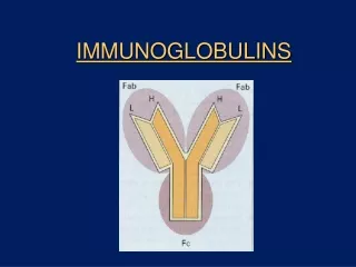
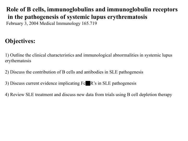
Role of B cells, immunoglobulins and immunoglobulin receptors in the pathogenesis of systemic lupus erythrematosis February 3, 2004 Medical Immunology 165.719. Objectives: 1) Outline the clinical characteristics and immunological abnormalities in systemic lupus erythematosis

E N D
Role of B cells, immunoglobulins and immunoglobulin receptors in the pathogenesis of systemic lupus erythrematosis February 3, 2004 Medical Immunology 165.719 Objectives: 1) Outline the clinical characteristics and immunological abnormalities in systemic lupus erythematosis 2) Discuss the contribution of B cells and antibodies in SLE pathogenesis 3) Discuss current evidence implicating FcgR’s in SLE pathogenesis 4) Review SLE treatment and discuss new data from trials using B cell depletion therapy
Systemic lupus erythematosus (SLE) Major clinical manifestations: -> Red rash or color change on the face, often in the shape of a butterfly across the nose and cheeks *** -> Painful or swollen joints -> Unexplained fever -> Chest pain with deep breathing -> Extreme fatigue -> Sensitivity to the sun -> Depression, trouble thinking, and/or memory problems Population affected: -> incidence around 1/2000 in North America -> 8:1 female/male -> higher incidence and severity in African American and Hispanic women
SLE: Major diagnostic laboratory findings: -> Positive lupus erythematosus cell preparation (a peculiar polymorphonuclear leukocyte which has injected nuclear material) -> Hemolytic anemia, leukopenia, lymphopenia or thrombocytopenia -> Heavy proteinuria or cellular casts in urine sediment -> ***Anti-nuclear and/or anti-DNA antibodies*** Related systemic autoimmune diseases with overlapping findings: -> Rheumatoid arthritis (mainly restricted to joints, distinguished by presence of RFs) -> Sjogren’s symdrome (mainly restricted to salivary and lacrimal glands) -> Schleroderma (mainly restricted to skin) LE cell 1948 ANA
ANA Fluorescence Patterns and Disease Association Nuclear Fluorescence Pattern Rim (peripheral) Homogeneous (diffuse) Speckled Nucleolar Disease Association SLE Drug-induced LE, SLE SLE, Sjogren's, scleroderma Scleroderma -> 95% of SLE patients are ANA positive, thus a negative result virtually excludes SLE -> but ANA can also found in other autoimmune diseases and chronic infections, thus a positive result cannot be the sole basis for diagnosis
Identity of specific molecular targets of ANAs ***Anti-double-stranded DNA -> gives rim staining pattern in ANA -> most important for SLE (2/3 of patients have + quite specific for SLE) Anti-single stranded DNA -> less specific Anti-histone antibodies Anti-ribonuclear proteins -> antibodies against protein-RNA complexes -> anti-Sm present in 1/3 of SLE patients, not in other conditions -> anti-U1-RNP, anti-SS-A/B
Immune complexes in SLE pathogenesis Mechanism for glomerulonephritis: Anti-DNA antibodies in SLE tend to have high isoelectric point (net positive charge) which promotes their binding to DNA Circulating DNA from damaged or dying cells can bind to the basement glomerular basement membrane Anti-DNA Abs binding to DNA on the basement membrane can fix complement Complement split products (C3a, C5a) trigger inflammatory response Lupus glomerulonephritis Similar mechanisms for skin lesions?
Other pathogenic roles of auto-Ab in SLE Anti-red cell and anti-platelets Abs -> hemolytic anemia, thrombocytopenia Anti-cardiolipin antibodies -> miscarriages, vascular thrombosis Anti-T cell antibodies -> immune dysfunction?
Immunological abnormalities in SLE B cell / antibody / complement systems: -> increased numbers of plasma cells in the bone marrow and peripheral lymphoid tissues -> limited repertoire of Ig genes used in autoantibodies -> antigen-driven-clonal expansion -> progressive accumulation of somatic mutations in Ig genes used in autoantibodies (affinity maturation?) -> marked accumulation of circulating immune complexes during acute flares -> decreased clearance of immune complexes through Fc receptors and complement receptors (cause or effect of increased IC?) -> decreased circulating C3/C4, increased split products such as C3a, C3d T cell system: -> in some cases, anti-T cell antibodies -> T cell lymphopenia -> generalized depression in cell mediated immunity -> apparent oligoclonal expansion of pathogenic T cells
Genetic factors in SLE -> different disease frequencies in different ethnic groups -> sibling recurrence-risk ratio ~15-20 (thus, if your sibling has SLE your risk is ~1% rather than ~0.05%) -> high clinical disease concordance among twins: 2-5% for dizygotic twins, 24-58% for monozygotic twins -> complement deficient patients (rare) very frequently develop SLE (40-75%) -> evidence for linkage to specific gene loci: Environmental factors?
Animal models for studying SLE genetics and pathogenesis (NZBxNZW)F1 -> Spontaneously develop a systemic autoimmune disease similar to lupus -> autoantibodies, immune complex disease, premature death -> Parental NZB mice have milder for of disease -> Useful model for dissecting the complex genetics of the disease -> multigenic, with different genes controlling different immunological abnormalities/production of specifc autoantibodies MRL lpr/lpr and MRLgld -> develop SLE-like syndrome associated with massive lymphoproliferation/splenomegaly -> gene defect identified as mutation in the death receptor Fas (lpr) or mutation in Fas ligand (gld)
Evidence implicating Fcg receptors in lupus -> Genetic deficiency of activation receptors attenuates disease in lupus-prone mouse strains -> Reduced expression of inhibitory receptors in autoimmune-prone mouse strains due to promotor polymorphisms -> Genetic deficiency of inhibitory receptor leads to spontaneous development of lupus in some non-lupus prone mouse strains -> Human linkage studies
Interaction between FcgRII and other genetic factors Bolland et al, J. Exp. Med 195: 1167
Effect of FcgRIIB retroviral-mediated bone marrow transduction on spontaneous autoimmunity in lupus-prone mice
Retroviral transduction of FcgRIIB inhibits kidney pathology
Retroviral transduction only increased FcgRII expression by 2-fold in about half of B cells! Small changes tip the balance Marked reduction in HAS-negative population: less B cell activation
FcgRII reguates the accumulation of autoreactive plasma cells through a cell-autonomous feedback loop? Fukuyama et al, Nature Immunol. 6:99 Dec.12, 2004
Experimental treatments Rituximab Treatment of SLE Current standard treatments = immunosuppression -> anti-inflammatories: corticosteroids, NSAIDs -> immunosuppressives: cyclophosphamide, prednisone, methotrexate, hydroxychloroquine, Azathioprine -> I.e. severe patients ore on some very nasty drugs
Efficacy of B cell depletion 11/17 patients showed effective depletion (some other studies have seen higher) Looney et al, Arthritis Rheum. 50: 2580
Improved Systemic Lupus Activity Measure (SLAM) scores, but no reduction in titre of anti-dsDNA -> Clinical response was especially notable for rashes, mucositis, alopecia, and arthritis -> The average daily dose of prednisone was 13 mg at the start of the study and 10 mg at the completion of the study -> Overall, rituximab was well tolerated. Only 1 mild infusion reaction was noted (bronchospasm)
Development of human anti-chimeric antibody (HACA) in some patients is associated with poor B cell depletion
Summary of B cell depletion therapy for SLE -> Rituximab holds significant promise for treatment of severe refractory SLE -> Clinical response is linked with B cell depletion; however, total Ig levels and anti-dsDNA levels are not markedly affected (why?? Selective targeting of sub-populations?, modulation Of costimulatory molecule expression?) -> therapy seems safe and well-tolerated -> more extensive controlled trials are warranted -> “Despite lack of marketing authorization, rituximab is already being used to treat various refractory autoimmune diseases in daily rheumatological practice” -> as for all monoclonal Ab therapies, efficacy might be improved with fully human Abs