Download
1 / 27
280 likes | 296 Views
IMMUNOGLOBULINS. Immunoglobulins. Humoral basis of immunity – 19 th century. Introduction of an Ag into an animal Abs appeared in the serum & body fluids Immune sera Abs react with Ag in a specific & observable manner. Distribution of Major Human Ig.

E N D
Immunoglobulins • Humoral basis of immunity – 19th century. Introduction of an Ag into an animal Abs appeared in the serum & body fluids Immune sera • Abs react with Ag in a specific & observable manner.
Distribution of Major Human Ig • Electrophoresis of human serum separated serum proteins into 2 major components : 1. Soluble Albumins 2. Insoluble Globulins - , & • Ab activity - γ globulin fraction of serum proteins. • 1964 – ‘Immunoglobulin’ by WHO.
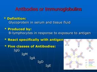
Igs are synthesised mainly by the plasma cells & to some extent by the lymphocytes. • Constitutes 20-25% of total serum proteins. • 5 classes of Igs – IgG, IgA, IgM, IgD & IgE.
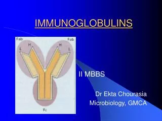
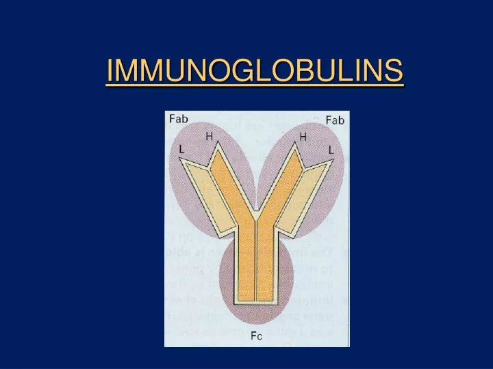
Structure of an Ig • Glycoproteins. • 2 pairs of polypeptide chains – 2 light (L) chains & 2 heavy (H) chains. • “L” chain – smaller chain - low molecular wt (25,000) • “H” chain – larger chain - high molecular wt (50,000)
Structure • L chain attached to H chain by disulphide & non- covalent bonds. • L chains : 2 forms – kappa () & lambda () • Each molecule of Ig can have either or , but never both. • H chains : structurally & antigenically distinct for each class.
H chain • H chain designated by Greek letter.
Structure • Light chain – 2 regions Constant (CL ) Variable (VL ) C – terminal N - terminal
Structure • H chain also divided into VH & CH regions; the CH region is further divided into CH1, CH2 & CH3. • Regions also called as DOMAINS : - globular in shape - stabilized by intrachain disulphide bonds • Ag binding sites are located in the variable domains.
Hypervariable regions • Amino acid sequence in the variable region of L & H chains are not uniformly variable. • Consists of some highly variable(hypervariable) & some relatively invariable zones. • Highly variable zones actually make contact with the epitope on an Ag and are called as Complementarity Determining Regions (CDRs) • 3 CDRs – each made up of 9 -12 amino acids. CDR3 is the longest & most variable of the three.
Structure • ‘ Hinge ’ region – segment of H chain between CH1 & CH2. • Flexibility to Ab • Susceptible to enzymes & chemicals. • Studies involving the cleavage of Ig molecule by pepsin & papain have led to a detailed picture of Ig structure.
Fragments of Ig • Fab – Ag binding. Fc fragment • Composed of carboxy terminal of H chain. • Determines the biological properties of Ig molecule. • Receptors for Fc portion expressed by – mononuclear cells neutrophils phagocytosis NK cells tumour cell killing eosinophils & mast cell degranulation mast cells
Ig G • Major serum Ig • Constitutes 75% of total Igs. • 4 subclasses found in humans – IgG1, IgG2, IgG3 & IgG4, each having a distinct type of gamma chain. • Major Ab of secondary response, found both in serum & body fluids. • Only maternal Ig to be transported across placenta – natural passive immunity in newborn. • Participates in complement fixation, precipitation & neutralisation of viruses & toxins.
Ig M • 5-8 % of serum Igs. • Short lived Abs. • Pentameric structure. • Predominant Ab in primary immune response. • Earliest Ab to be synthesized by the fetus. • Confined to the intravascular pool due to its large size. • Not transported across placenta. • Presence of IgM in newborn indicates intra uterine infection. • Useful in the diagnosis of congenital infections like syphilis, rubella, HIV, toxoplasmosis etc.
Ig A • 2nd most abundant, constitutes 10-13 %. • Major Ig in the colostrum, saliva, tears & other body fluids. • Occur in 2 forms : IgA1 & IgA2. • Secretory IgA is always in dimeric form – composed of 2 basic chain units, a J chain & the secretory component. • Secretory component helps to transport the dimer from the submucosa to the mucosal cell surface. • Secretory component protects IgA from proteolytic digestion and denaturation.
Ig D • Resembles Ig G structurally. • Occurs along with Ig M on the surface of B cells • Very susceptible to proteolytic attack.
Ig E • Present in very low levels in serum. • Found on the surface of mast cells & basophils which have specific receptors for the Fc portion of IgE. • Chiefly produced in the linings of respiratory & intestinal tracts. • Responsible for anaphylactic type of hypersensitivity. • Defense against parasitic infections.
Abnormal Igs • Structurally similar proteins in serum seen in certain pathological conditions. • Bence Jones protein in multiple myeloma – light chains of Igs. • Cryoglobulinemia – formation of gel or ppt on cooling the serum which redissolves on warming – in myelomas, SLE etc.
Antibody diversity • An individual produces a large number of Abs to cope with the vast number of different Ags. • This Ab diversity is due to the Ig genes. • Genes coding for the variable & constant portions of the chains are separate • One or only few genes code for C region whereas many genes code for the V region.
Generation of diversity • Multiple V- region genes. • V-J & V-D-J recombination. • Junctional diversity • Nucleotide addition – extra nucleotides may get inserted between VH & D, and between D & JH segments • Somatic mutation – point mutation in the genes for V domain.