Download
1 / 44
480 likes | 984 Views
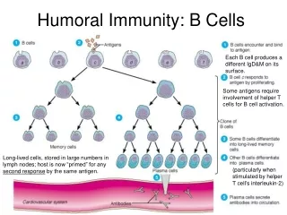
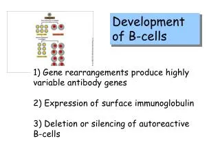
Development of B-cells. 1) Gene rearrangements produce highly variable antibody genes 2) Expression of surface immunoglobulin 3) Deletion or silencing of autoreactive B-cells. allelic exclusion.

E N D
Development of B-cells 1) Gene rearrangements produce highly variable antibody genes 2) Expression of surface immunoglobulin 3) Deletion or silencing of autoreactive B-cells
allelic exclusion Allelic exclusion is the process by which lymphocytes express antigen receptors from only one of two possible alleles.
Membrane bound antigens can crosslink the surface IgM on the surface of immature B cells. Examples of such antigens are MHC class I and class II molecules. Immature B cells that recognize such self antigens undergo apoptosis and are therefore removed from the repertoire.
Stem cell Germline DNA Early pro-B cell Late pro-B cell Pre-B cell IgM expressed on cell surface Immature B cell Mature B cell Secreted Ig Plasma cell
Four sources of antibody diversity: 1) Combinatorial diversity 2) Diversity due to heavy and light chain joining 3) Junctional diversity 4) Diversity due to somatic mutations
Combinatorial Diversification In the mouse variable light chain, about 300 different V-regions can be joined to 4 different J segments which are then joined to one C-region. In the mouse variable heavy chain, 500 different V-regions can be joined to 4 J segments and 12 D segments. What is the total number of possible combinations?
Combinatorial Diversification Light Variable Chain: 300 x 4 = 1200 Heavy Variable Chain: 500 x 4 x 12 = 24,000 Total possible combinations: 1200 x 24,000 = 28,800,000
Junctional Diversification Joining of the V segment to a J or D segment is not always precise in that random nucleotides can be inserted or lost at the junction. It is estimated that junctional diversification increases the number of possible combinations by at least 108.
VDJ recombination involves several steps, beginning with a cut to the DNA. These site-specific double- stranded breaks are initiated by two genes-called RAG-1 and RAG-2.
Mechanisms of variable region DNA rearrangements • Recombination signal sequences (RSS's) • Recombination activating genes (RAG1 and RAG2)
A protein, called Ku80, appears to do double duty in the processes of DNA repair and a kind of DNA recombination known as "VDJ joining."
VDJ recombination also takes place in the T cells. In this case the product is the TCR (T cell receptor protein).
Class Switch In immature (and naïve) B cells, the m (and d) chains are located adjacent to the VDJ region. At this point, IgM (and IgD) are expressed. Constant region gene segments located further downstream can be brought into proximity to the VDJ region by looping out and deleting intervening sequences. This process is irreversible and requires T cell signals.
B cell derived tumors Chronic lymphocytic leukemia Blood Malignant Plasma Cells Multiple myeloma Plasma cell (secreting Ig of various isotypes, IgG, IgA, IgE...)
Monoclonal Antibodies A hybridoma is a fusion of a cancer cell with a functional, normal cell - a hybrid of a lymphoma) which results in the generation of an immortal cell-line.
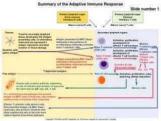
T Cells and Lymphokines T cells contribute to the immune defenses in two major ways. Regulatory T cells are vital to orchestrating the elaborate system. (B cells, for instance, cannot make antibody against most substances without T cell help). Cytotoxic T cells, on the other hand, directly attack body cells that are infected or malignant.
Immune System Cell Types are Defined by Cell-specific CD antigens. Found on thymocyte subsets, helper and inflammatory T cells (about two thirds of peripheral T cells), monocytes, macrophages. CD4 CD4 acts as a co-receptor for MHC class II molecules and as a receptor for HIV-I and HIV-2 gp120.
CD4+ cells are also known as helper Tlymphocytes, while CD8+ cells are known as cytotoxic T lymphocytes.
Each CTL (Cytotoxic T-lymphocyte) (CD8+) has a unique surface molecule much like an immunoglobulin, called a T-Cell Receptor (TCR).
TCR’s are restricted to only being able to recognize short amino acid chains (epitopes), which are displayed on the surface of cells in conjunction with a particular Major Histocompatibility Complex protein (MHC). MHC Class II HLA-DR1
Analysis of the human beta T-cell receptor (TCR) locus comprising a complex family of genes was reported in the June 21, 1996, issue of Science (272, 1755-62).
Almost all the cells constantly produce MHC proteins and attach small internal proteins to it for expression on their surface. CTLs probe the surface of cells for MHC-small protein complexes.
Class I genes are present on nearly all cells, and encode glycoproteinson the surface. These glycoproteins change when abnormal changes occur in the cell, thereby signalling cytotoxic T cells of malfunction within the cell. Class I genes are important in the identification of tumor cells, and cells that have been taken over by viruses.
Class II genes encode glycoproteins on the surface of antigen presenting cells, such as macrophages and B cells. These glycoproteins are used to present recognizable portions of antigens for use by helper T cells to form a global immune response.
In most cases, as with B-Cells, the Cytotoxic T-Cells (CD8+) never finds a complex which its unique TCR recognizes. However, in the event that it does, it grows and divides into mature CTLs and memory CTLs.
Helper T-Cells produce chemical factors that regulate all other aspects of the Immune System. Like the CD8+ cells, the TCR proteins on helper T-cells act by recognizing peptides bound to self MHC class II molecules.
"interleukins” -messengers between leukocytes IL-2 is produced by T cells and has potent effects on the maturation and proliferation of a number of cell types, including T-cell, B-cells and natural killer cells. IL-2 is produced as a recombinant DNA drug product and is licensed in the United States for the treatment of patients with metastatic cancers.
Helper T cell begin the attack by binding to the macrophage via the T cell's antigen receptor. This union, aided by a third cell (dendritic cell) that makes immune cells cluster, stimulates both the macrophage and the helper T cell to exchange chemical messages between themselves and with other cells of the immune system.
The macrophage releases cytokines called interleukin-1 (IL-1) and TNF, or tumor necrosis factor. TNF steps up the production of IL-1 and performs many of the same functions that IL-1 does, including inducing fever to help the body combat infection more effectively. IL-1 alone enhances the ability of dendritic cells to form immune cell clusters and stimulates the helper T cell to release several lymphokines, one of which is called interleukin-2 (IL-2).
IL-2 in turn causes T cells to secrete gamma interferon, which, among other things, activates macrophages. IL-2 also instructs other helper T cells and a different class of T cells, the killer T cells, to multiply.