Download
1 / 44
440 likes | 553 Views
Muscle tissues. Smooth m. Cardiac m. Skeletal m. Organisation of skeletal muscles. A muscle consists of many m. fibers It is covered by epimysium The fibers are collected into fascicles Each fascicle is coverd by perimysium. The individual m.fiber is coverd by endomysium

E N D
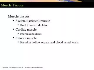
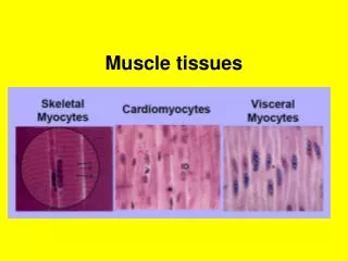
Muscle tissues Smooth m. Cardiac m. Skeletal m.
Organisation of skeletal muscles • A muscle consists of many m. fibers • It is covered by epimysium • The fibers are collected into fascicles • Each fascicle is coverd by perimysium
The individual m.fiber is coverd by endomysium • The CT of the muscle blend together at each end to form the tendon which scrues the m. to bones
Skeletal muscles The muscles contract in response to motor nerve impulses arriving at neuromuscular junction.
The major functions of sk. M. • Force production for locomotion & breathing • Force production formaintaining posture & stabilizing joints • Heat production • Helps venous drainage
Contraction • Transmission of action potential at neuromuscular junction by neurotransmitterAcetylcholine • Spread of action potential along the sarcolemma & penetrates deep into T. tubules • Release of calcium from Sarcoplasmic reticulum
Calcium binds troponin C causes tropomyosin to move away from its position covering the actin active sites • Binding of myosin to actin filamints forming Cross-bridges
ATP provides energy for sliding of the 2 filaments • Shortening of sarcomere occurs
The muscle then relaxes when calcium is pumped back into SR. This breaks the cross- bridges. Then they slide back & lengthening occurs
Principle skeletal muscles • Muscles of the face & neck • Muscles of the trunk • Mucsles of the shoulder & upper limb • Mucsles of the hip & lower limb
Muscles of the face • Occipitofrontalis: it raises the eyebrows. • Levator palpebrae superioris: raise the eyelid. • Orbicularis oculi:closes the eyes
Orbicularis oris:colsesthe lips • Muscles of Mastication: • Masseter • Temporalis • Pterygoid
Muscles of the Neck • Sternocleidomastoid:when thew muscle contracts on one side it drws the head towrds the shoulder.when both contracts they flex the neck • Trapezius: pulls the head bach wards & controls movement of scapula
Muscles of the back • Trapezius • Latissimus dorsi • Teres major • Poas • Quadratus lumborum • Sacrospinalis
Muscles of abdominal wall • Rectus abdominis • External oblique • Internal oblique • Trasversus abdominis • Quadratus lumborum
Functions • Compress the abdominal organs • Flex the vertebral column in lumbar region
Muscles of pelvic floor • Levator ani • Coccygeus They support the pelvic organs & maintain continence
Mucsles of the shoulder & upper limb • Deltoid: ant. flexion middle abduction post. extension • Pectoralis : flex & adduct • Coracobrachialis: flex
Biceps: short head Long head Distal tendon Action: on shoulder? on elbow ? • Brachialis: main flexor of elbow
Triceps: 3 heads? Action: Shoulder? elbow? • Brachioradialis: flex elbow • Pronator quadratus: pronation of hand • Pronator teres: pronation of hand • Supinator : supination
Flexor carpi radialis • Flexor carpi ulnaris • Extensor carpi radialis • Extensor carpi ulnaris • Palmaris longus • Extensor digitorum
Muscles that control finger movement • Large m. in the forearm that extend to the hand • Smaller m. which originate from carpal & metacarpal bones ( Thenar & hypothenar m.)
Muscles of the hip & lower limb • Psoas: flex the hip • Iliacus: flex the hip • Quadriceps femoris • Obturators: lateral rotation of hip • Gluteals: extension. abd. & med. rotation of hip
Sartorius: 2 actions? • Adductor group • Hamstrings • Gastrocnemius:2actions? • Anterior tibialis: dorsi flexion of foot • Soleus: plantar flexion of ankle