Download

1 / 37
900 likes | 3.24k Views
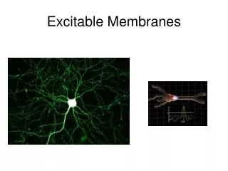
The excitable tissues (Nerve+ Muscle). The nerve. Neuron:- - DIF;- unit of function of the central nervous system Parts of motor neuron & function of each part : 1- Soma (cell body) 2-Dendrites carry nerve impulses from surroundings to the soma

E N D
Neuron:- -DIF;-unit of function of the central nervous system Parts of motor neuron & function of each part: 1- Soma (cell body) 2-Dendrites carrynerve impulses from surroundings to the soma 3 Axon hillock at which nerve impulses begin 4-Axon & axon terminal
-Histological classification of axons:- 1- myelinated : have myelin sheath (diameter more than 1um) 2- unmyelinated (diameter less than1um ) -type C:postganglionic autonomic &pain fibers
-Myelin sheath is formed by schwann cell which deposit sphingomyelin Functions of myelin sheath 1-insulator 3- increase conduction velocity
RESTING MEMBRANE POTENTIAL DIF:- it is potential difference across membrane during rest (without stimulation) Value:- -70 to-90 mv in large nerve fibers ( -ve inside) - -The membrane is polarized
CAUSES/ 1- Contribution of K & Na diffusion potential through Na & K leak channels of nerve membrane, CM is more permeable to K than to Na, thus K tends to leak to the out side (down its concentration gradient) carrying positive charge with it. This make the cell interior more negative 2-Active transport of Na & K ions( Na/K pump) 3- Negative ions inside membrane as phosphate & proteins
Causes of RMP: 1. RMP is 100 times more permeable to K+ than Na+. K+ tends to leak out of the cell down its conc gradient, carrying +ve charge with it. (through K leak channels). 2. non-diffusible anions (proteins, sulphate and phosphate ions) cannot leave the cell. 3. very small amount of Na+ diffuses into the cell down its conc gradient. The mb only slightly permeable to Na+. (through Na+ leak channels). 4. Na+-K+ pump maintain conc gradients of K+, and Na+ between the two sides of the mb.
Origin of RMP: 1- Contribution of K diffusion potential:- N.B/ K diffusion contributes far more to membrane potential . -K leak channels:- K OUTFLUX TO OUTSIDE causing –ve inside (from high conc inside to outside carrying +ve charge with it→ electropositivity outside& electronegativity inside
What does it mean when a neuron “fires”? • Firing = excitability = action potential = nerve impulse • Recall resting potential of all cells • High K+ in; high Na+ out • Cell is polarized • Cell overall neg. charge inside due to molecules like proteins, RNA, DNA • Charge measured in millivolts • Potential = difference in charge across PM • Current = flow of charge (ions) from one point to another
2- Contribution of Na diffusion potential:- • Na leak channels :- Slight membrane permeability to Na ions in leak channels from outside to inside.(why slight?) • 3. Na+-K+ pump maintain conc gradients of K+, and Na+ between the two sides of the mb.
Changes that occure through the nerve after stimulation by threshold (effective) stimulus:- 1- Electrical changes (nerve action potential) 2- Excitability changes 3-Thermal changes 4-Chemical changes
The action potential • It is sudden reversal of membrane polarity produced by a stimulus to produce a physiological effect such as: • Transmission of impulse along nerve fibres • Release of neurotransmitters • Muscle contraction • Activation or inhibition of glandular secretion
1- Electrical changes The nerve action potential -It is potential difference along nerve membrane after stimulation by threshold (effective)stimulus -oscilloscope to measure rapid changes in membrane potential -Nerve signals (impulses) are transmitted as nerve action potentials conducted along the nerve fiber as a wave of depolarization to its end -The factors necessary for nerve action potential arevoltage gated Na & k channels -
Summary of events that causes AP:- 1-Initiation of Action Potential (AP) - -70 to-90 mv is the resting potential - Threshold stimulus open voltage gated Na channels & Na influx rises resting potential from -90 towards zero (gradual depolarization) -as membrane potential raises ---------- open more Na channels & more Na influx (+ve feedback ) until all voltage gated Na channels open.
2-Depolarization occurs & membrane potential reach zero value to reach + 35 mv, -at + 35 mv all Na channels begin to close suddenly( Depolarization ends)
c-Repolarization:- due to high K conductance( flow) to outside (K outflux) by openning of all voltage gated K channels (causes negativity inside -
Hyperpolarization: Why? • Na-K pump now start to move Na out & K in against their concentration gradient, so the RMP is resumed and the membrane is ready for another stimulus
The action potential (cont.)*** Threshold stimulus: If a stimulus is strong enough to move RMP from its resting value (-70mV) to the level of (-55mV) which leads to production of an AP
Subthreshold stimulus Stimulus that result only in local depolarisation
All or nothing principle:- - Once threshold value for excitation is reached a full AP produced ,its intensity can not increased by increasing stimulus intensity ( suprathreshold) Direction of propagation of AP:-in one direction
What happens after an action potential? • Refractory period: few millisecs • Time during which can’t stimulate neuron a second time • Happens until recovery of resting potential • Two stages • Absolute refractory period • No new action potential possible • Relative refractory period • Can trigger new action potential if stimulus is very strong
Propagation of action potential( 1- in myelinated nerve fibers:- Saltatory conduction ( jumping) Value:- 1-↑ velocity of conduction of nerve impulses) 2-Conserve energy for axon because only nodes depolarize
How do action potentials travel down the axon? • Myelinated sheaths • Many times faster transmission • Action potential skips from one node of Ranvier to the next • Called saltatory conduction • http://www.blackwellpublishing.com/matthews/actionp.html
2- Non- myelinated nerves:- (local circuits)=point to point -depolarization pass by local circuits. -
What else influences speed of action potential? .Axon diameter -The larger the diameter, the faster the speed of transmission -Less resistance to current flow with larger diameter Slower transduction Faster transduction
What happens if myelination is lost? • Multiple sclerosis • Autoimmune disease • Usually young adults • Blindness, problems controlling muscles • Ultimately paralysis • Immune system attacks myelin sheaths and nerve fibers • Scar tissue (scleroses) replaces some damaged cells • Other now unmyelinated axons sprout Na+ channels • Accounts for sporadic nature of disease?