Download
1 / 27
270 likes | 404 Views
Cancer Stem Cells, Clonal Heterogeneity and Clonal Tides in Multiple Myeloma. Linda M. Pilarski University of Alberta, Canada 23 October 2014. To Monitor the Myeloma Clone and Response to Treatment:.

E N D
Cancer Stem Cells, Clonal Heterogeneity and Clonal Tides in Multiple Myeloma Linda M. Pilarski University of Alberta, Canada 23 October 2014
To Monitor the Myeloma Clone and Response to Treatment: Essential to change the focus from screening only the plasma cell (PC) compartment of MM to looking at the entirety of a MM clone Each MM clone includes PC, B cells and probably a CD20+ CD34+ cancer stem cell compartment
Each MM clone has multiple phenotypically defined compartments that harbor the clonotypicIgH “wolves in sheep’s clothing”
MM Cancer Stem Cell Function is Multi-Faceted and Influenced by Parameters of Maturation and Time • Early stage progenitors: Extensive self renewal, prolonged time to detectable disease • Late stage progenitors: Limited self renewal, rapid onset of disease symptoms
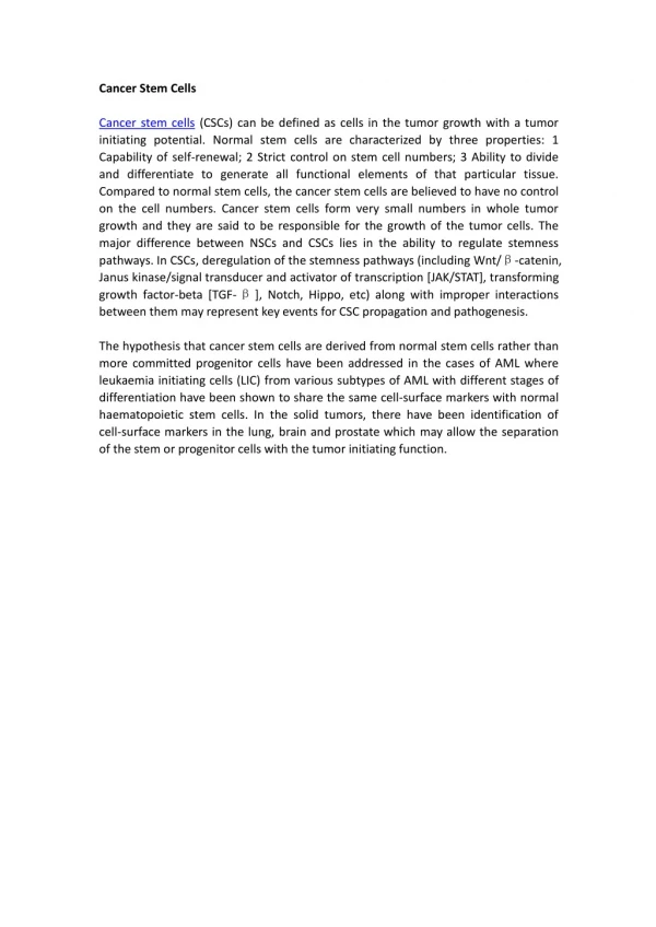
Defining a Cancer Stem Cell • 1.Able to generate/regenerate • the malignant clone • Exhibits self renewal • 3. Quiescent and drug resistant • 4. In MM, may be a complex • population comprised of • multiple differentiation stages • MM remains incurable. This must mean • that current MM therapies fail to kill • MM stem cells
MM BM lymphs have chromosomal abnormalities 17 “chromotypes” in BM lymphsfrom ~45% of MM Pts. 2-37% of total BM lymphocytes harbor abnormalities DebesMarun et al, 2012
MM stem cell capability appears to reside in both B and plasma cell (PC) compartments “Early stage” chromosomal abnormalities (IgH translocations, del13) appear to arise in B cells and are transmitted to PC as MM clonal expansion and differentiation occurs Late abnormalities associated with disease progression (del17p and amp1q21) occur only in PC. This suggests that as disease progresses, PC may acquire some degree of generative capability
Chromosomally abnormal BM lymphocytes correlate with reduced survival in MM MM patients with abnormalities in both PC+ lymphshave significantly worse survival compared to those with abnormalities only in PC (p=0.016) DebesMarun et al, 2012
MM cancer stem cells (MM-CSC): Clonotypic CD20+ MM-CSC (B cells) show self renewal, proliferative quiescence, drug resistance and the ability to regenerate disease P1 = 100% B cells P1 and P3 = 100% clonotypic Control P6 - B cells P6- Plasma cells Kirshner et al, Blood 2008
MM Cancer Stem Cells Co-purify with Hematopoietic Progenitor Populations CD34-purified cells from MM mobilized blood autografts have clonotypic transcripts (mean = 31% of CD34+45lo cells) CD34-purified cells from MM autografts include DNA aneuploid cells (mean= 31% hyperdiploid) CD34-enriched cells from MM autografts engraft human MM to NOD SCID mice (from 6/7 mobilized bloods) CD34-enriched cells from MM autografts cause MM bone lesions in 76% of xenografted NOD SCID mice Pilarski and Belch 2002
The myeloma clone is a dominant clone • Dominant B cell clones prevent expansion • of other clones • Other clones arise with loss of dominance • Speculation and Questions: • Transformation events may be frequent in MM • Does MM dominance prevent expansion of • other potentially malignant clones? • Do “submissive” clones become progenitors • for second cancers?
Myeloma is characterized by “clonal tides” (reviewed by Bahlis, Blood 2012) 1. Over time, clonal waves expand and contract 2. The assumption is that these represent variants of the MM clone with acquired genomic changes Tidal waves share some common abnormalities 3. No direct evidence to confirm that all tidal waves arise from the same MM “parent”. 4. Must be confirmed through IgH VDJ analysis
Dual clones occur frequently in MM • 1. 20% of MM patients have a second clone • (distinct IgHVDJ) • 2. Second clones are frequent (0.01-40% of MNC) • 3. Second clones harbor genetic abnormalities • 4. Second clones persist throughout treatment • Does treatment stimulate second clones and/or • allow submissive clones to escape MM dominance? • Do second clones form a pool of progenitors that • can lead to second cancers? Kriangkum et al. 2013
Dual clones have different IgH VDJ, geographic locations, frequencies and chromosomal abnormalities (12/46 patients) Patient #6 del13 amp1q21 T(11;14) Clone 1Clone 2 Sternal biopsy Iliac biopsies CDR3 = 63bp CDR3 = 42 bp VH4-30 VH1-18 91% of PC 67% of PC Productive IgH Productive IgH
Clone 1 and Clone 2 are in different cells (single cell analysis) C1 = 100% of BM PC C2 = 10% of BM B cells C2 = 25% of PB B cells C1 = 60% of BM PC C2 = 19% of BM B cells C2 = 10% of PB B cells P/NP BiallelicIgH rearrangements
Expansion of a Second Clone After Treatment, at Relapse Treated for 6 years with thalidomide Diagnosis to year 2 - C2 at low frequency in PB, dormant? Years 2-7 - extensive clonal expansion of C2 in BM
Frequencies of MM clone and Second Clones in Genomic DNA of MNC for 12 MM patients (single cell PCR and/or real time quantitative PCR) BM Blood Clone 1 (MM) 0.1-70% 0.01-41% median = 30% median = 0.5% Clone 2 0.06-24% 0.1-31% median = 2.5% median = 1.0% Values = % of mononuclear cells
IgH Clonal Diversity in MM Pts with Two Clones, Measured by ImmunoSEQ ~100,000 to >1 million reads C1 C2 C1 C2 ImmSEQ 74% 25% 84% 9% Q-PCR 37% 24% 30% 3% Patient 1 Patient 2 Uni-clonal MM Massively Parallel Sequencing by Adaptive Biotechnologies
The frequency of “second” clones is such that they would likely score as distinct clones in array analysis or whole genome sequencing. Must distinguish between : intra-clonal diversity (related clones) and inter-clonal diversity (unrelated clones). Clonal dynamics in MM involve B lineage expansions So…… IgH VDJ gene rearrangements are the ONLY unequivocal definition of relatedness.
Assumptions of commonality/relatedness of tidal clones are based on the presence of shared chromosomal abnormalities e.g. insertions and deletions identified by CGH Is this sufficient?
Do chromosomal abnormalities arise prior to IgH VDJ rearrangement? If so, post-IgH rearrangement clonal dynamics may reflect unrelated and clonally distinct transforming events. (inter-clonal diversity) Shared abnormalities could be present in multiple “parent” B cells, leading to independent clonal expansions and discrete clonal tides Clonal tides may include both intra-clonal and inter-clonal diversity
TIME Hematopoietic Progenitors and/or Pro-B cells Copy Number Aberrations/Aneuploidy IgH VDJ Rearrangement VDJ-1 VDJ-2 VDJ-3 VDJ-4 more …. Transforming Events & Clonal Dynamics Dominant MM clone
Inter- and Intra- clonal Tides: Dynamics of Independent Clones in MM Clone VDJ1 Dominant MM clone Clone VDJ2/Intra-clonal tides IgH VDJ Gene rearrangement Clone VDJ3 Escape from dominance Clone VDJ4 TIME
Clonal dynamics may involve multiple discrete clones as well as multiple subclones • Related clones arise from the same B cell • - may acquire additional genetic aberrations • - clones will share the same IgH VDJ • - intra-clonal diversity • and/or • Unrelated clones derive from different B cells • - may arise from a shared pre/pro B expansion • - clones have different IgH VDH rearrangements • - clones may have the same genetic abnormalities • - inter-clonal diversity • - clinical significance ? second malignancies?
Important to Consider the Potential Impact of Treatment on Clonal Dynamics Compromise dominance of the primary MM clone and/or compromise the MM niche to allow alternate clonal expansions 2. Preferentially deplete some clones but spare others Preferentially stimulate/inhibit clones or subclones The impact of treatment may differ in its effect on families of related clones as compared to that on collections of unrelated clones
Conclusions • MM cancer stem cells are resting B lymphocytes • with clonotypicIgH rearrangements • MM BM B-cells carry the same chromosomal • abnormalities as MM plasma cells, and contribute • to poor survival • 3. MM patients can harbor two dominant clones • single cell analysis & next-gen sequencing 4. MM may undergo multiple transformation events - unrelated clones may emerge when clonal dominance wanes Inter-clonal diversity may contribute to clonal tides and possibly to second lymphoid malignancies
Acknowledgments: Carina DebesMarun JitraKriangkum, PhD Andrew R. Belch, MD Christopher Venner, MD Thanks to the myeloma patients who so generously donate their BM and blood Alberta Cancer Foundation and Myeloma Alberta